Table of Contents
- Introduction
- Immunohistochemistry Workflow
- Immunohistochemistry Selection Guide
- Pioneering in IHC Technology
- Choosing a Detection System
- Avidin-Biotin Complex (ABC)-Based Detection
- VECTASTAIN® ABC Systems
- VECTASTAIN® ABC Kits
- Choosing a VECTASTAIN® ABC Kit
- > Customizing your VECTASTAIN® ABC Kit
- Polymer-Based Detection
- ImmPRESS® One-Step Polymer Systems
- > ImmPRESS® Two-Step Amplified Polymer Systems
- > Choosing an ImmPRESS® Polymer Kit
- > Multiple Antigen Labeling Simplified
- Species on Species Detection
- M.O.M.® (Mouse on Mouse) Immunodetection Kits
- Choosing an Enzyme Substrate
- Enzyme Substrates
- > Enzyme Substrate Properties
- Multiple Antigen Labeling
- Enzyme Substrate Combinations
- Counterstaining
- > Counterstain/Substrate Compatibility Table
- Blocking Background Signal
- Secondary and Tertiary Detection Reagents
- Mounting Media
- Accessory Reagents
Counterstaining
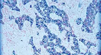
A counterstain introduces color to specific cellular structures to provide contrast to the colored enzyme substrate. Counterstaining aids in visualization and target localization, facilitating interpretation of morphology and cell structure within the tissue section. Our nuclear counterstains are packaged as convenient, ready-to-use solutions for use on individual slides or in staining dishes.
- Based on Gill’s III formulation
- Progressive stain formula. The intensity can be adjusted to optimize results for either manual or automated systems
- Excellent color contrast with most commonly used peroxidase and alkaline phosphatase substrates
- Suitable for use with non-aqueous and aqueous mounting media
- Alcohol- and mercury-free
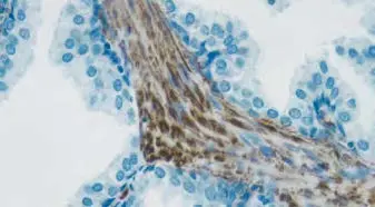
- Modification of Mayer’s hematoxylin developed especially forimmunocytochemistry
- Ready-to-use without filtration or ‘blueing’ step
- Stains in less than 45 seconds
- Excellent color contrast with most commonly used peroxidase and alkaline phosphatase substrates
- Suitable for use with non-aqueous and aqueous mounting media
- Mercury-free
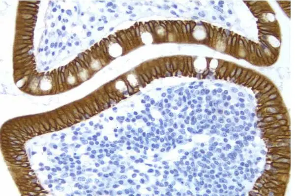
- Superior formulation of methyl green suitable for use with a wide range of enzyme substrates
- Simple, two step procedure
- Excellent alternative in multiple antigen labeling when hematoxylin obscures the substrate colors
- Suitable for use with non-aqueous mounting media
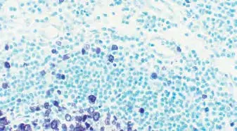
Vector Nuclear Fast Red (pink)
- Fast one-step protocol
- Excellent alternative in multiple antigen labeling when hematoxylin obscures the substrate colors
- Good contrast with a variety of substrates
