Table of Contents
- Introduction
- Immunohistochemistry Workflow
- Immunohistochemistry Selection Guide
- Pioneering in IHC Technology
- Choosing a Detection System
- Avidin-Biotin Complex (ABC)-Based Detection
- VECTASTAIN® ABC Systems
- VECTASTAIN® ABC Kits
- Choosing a VECTASTAIN® ABC Kit
- > Customizing your VECTASTAIN® ABC Kit
- Polymer-Based Detection
- ImmPRESS® One-Step Polymer Systems
- > ImmPRESS® Two-Step Amplified Polymer Systems
- > Choosing an ImmPRESS® Polymer Kit
- > Multiple Antigen Labeling Simplified
- Species on Species Detection
- M.O.M.® (Mouse on Mouse) Immunodetection Kits
- Choosing an Enzyme Substrate
- Enzyme Substrates
- > Enzyme Substrate Properties
- Multiple Antigen Labeling
- Enzyme Substrate Combinations
- Counterstaining
- > Counterstain/Substrate Compatibility Table
- Blocking Background Signal
- Secondary and Tertiary Detection Reagents
- Mounting Media
- Accessory Reagents
Pioneering in IHC Technology
Observation is one of the fundamental steps in the scientific method. However, for centuries the scientific study of tissues was limited to observations of dissections with the unaided eye (gross anatomy).
This all changed in the 17th century when Anton Van Leeuwenhoek fabricated a microscope that allowed observations of tissues at the cellular level, thus establishing the science of histology. While early researchers found it relatively simple to distinguish between the cell boundaries and subcellular compartments in plants, doing so in animal tissue presented a much greater challenge. It wasn’t until the late 19th century with the introduction of dyes, such as hematoxylin that Paul Mayer used to successfully stain nuclei, that the subcellular structure of tissues became visible and the science of histochemistry emerged.
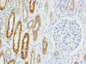
The number of available tissue dyes and stains increased during the early 20th century, as did the number of molecular families they identified. However, the ability to identify individual cellular- or tissue-specific proteins remained elusive. This changed in the mid-20th century when Dr. Albert Coons demonstrated that fluorescently labeled antibodies could be used to localize bacteria inside macrophages, thus pioneering IHC technology. Over the next two decades our understanding of antibodies, antigens and immunology grew rapidly. However, IHC remained largely a specialized research tool used primarily in university settings. Then in the late 1960’s, Dr. Stratis Avrameas and Dr. Paul Nakane independently developed methods to covalently couple the enzyme horseradish peroxidase (HRP) to antibodies. HRP in the presence of diaminobenzidine and hydrogen peroxide creates a brown precipitate at the site of the HRP-labeled antibody. The precipitate can be visualized using an ordinary light microscope. This allowed for the IHC results to be viewed in any lab having a light microscope, with no need for expensive, complicated fluorescence instrumentation.
The use of IHC as a research tool grew dramatically over the next decade. The technique began to be used in clinical settings at large university hospitals. The HRP assay system was further improved in the early 1980’s when Dr. Su-Ming Hsu showed that the high affinity of avidin for biotin could be used to increase the stability of the enzyme antibody complex and improve the sensitivity of the assay. Vector Laboratories was instrumental in the development and commercialization of IHC technology. The use of avidin- and biotin-based detection systems dominated the IHC market for the next two decades.
During this time, Dr. Shan-Rong Shi introduced “antigen retrieval” for formaldehyde-fixed tissues. This technique allowed IHC to be readily performed on formalin-fixed, paraffin-embedded tissues, greatly increasing clinical utility of IHC. However, in addition to improving antigenicity in tissue sections, antigen retrieval also exposed numerous sites of endogenous biotin that were previously undetected. This required steps to be added to IHC staining protocols to block endogenous biotin in biotin-containing specimens. In clinical settings in particular, antibody detection strategies returned to non-biotin HRP systems to avoid confusion resulting from endogenous biotin. However, the choice was not clear-cut, as avidin-biotin detection systems offered greater sensitivity than the previous peroxidase-labeled antibody systems.
This dilemma was finally resolved in the mid-2000s by the emergence of biotin-free polymer/multimer detection systems that offered similar sensitivity to avidin-biotin detection systems. Although early polymer-based systems suffered from background and tissue penetration problems, today’s IHC technology delivers performance comparable to the best avidin-biotin detection systems.
40-Year History of Quality and Innovation
1981
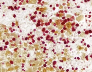
S.M. Hsu published that the high affinity of avidin for biotin could be used to increase the stability of the enzyme antibody complex
1993
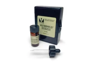
Vector Labs introduced first antifade mounting media for fluorescence (VECTASHIELD® Mounting Media)
1999
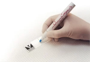
Vector Labs introduced a next-generation PAP pen conforming with environmental regulations (ImmEdge® Pen)
2000
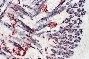
Vector Labs introduces M.O.M.® detection kits expanding the use of unconjugated mouse primary antibodies on mouse tissue sections.
2004
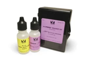
Vector Labs expanded antifade mounting media with hardening formulation (VECTASHIELD® HardSet™ Mounting Media)
2005
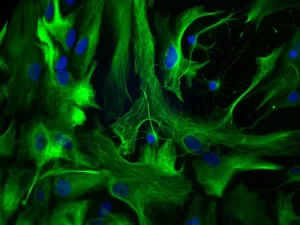
Vector Labs developed detection reagents enabling greater access to antigens within tissues and improving multiple antigen labeling (ImmPRESS® enzyme polymer)
2007
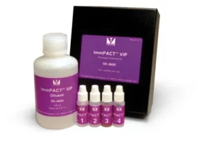
Vector Labs introduced proprietary substrates enabling multiplexing IHC (ImmPACT® HRP substrates)
2018

Vector Labs achieves ISO 9001:2015 certification
VECTASHIELD® Vibrance Antifade Mounting Media introduced
2020
Vector Labs adds SoluLINK Bioconjugation Product Line
VECTASHIELD® PLUS and H.O.H.™ Kit introduced
2021

Appointment of Lisa V. Sellers, Ph.D., as CEO. Strategic Investment by Thompson Street Capital Partners
VectaMount® Express Mounting Medium introduced
References:
1. Coons AH, Creech HJ and Jones RN “Immunological properties of an antibody containing a fluorescent group” Proc. Soc. Exp. Biol. Med. 47, 200-202 (1941)
2. Nakane P and Pierce GB Jr “Enzyme-labeled antibodies for the light and electron microscopic localization of tissue antigens” J. Cell. Biol. 33, 307-318 (1967)
3. Leduc E, Avrameas S and Bouteille M “Ultrastructural localization of antibody in differentiating plasma cells” J. Exp. Med. 127, 109-118. (1968)
4. Hsu S-M, Raine L, and Fanger H “Use of Avidin-Biotin-Peroxidase Complex (ABC) in Immunoperoxidase Techniques: A Comparison between ABC and Unlabeled Antibody (PAP) Procedures” J. Histochem. Cytochem. 29(4), 577-580 (1981)
5. Shi SR, Key ME and Kalra KL “Antigen retrieval in formalin-fixed, paraffinembedded tissues: an enhancement method for immunohistochemical staining based on microwave oven heating of tissue sections” J Histochem Cytochem. Jun, 39(6), 741-8 (1991)
6. Childs GV “History of Immunohistochemistry” Pathobiology of Human Disease. 3775-3796 (2014)

