In Situ Hybridization Workflow
Purification, characterization, and identification of nucleic acids (DNA or RNA) often require analysis of the target in blotting applications. Nucleic acids are usually separated by size using gel electrophoresis and transferred to a membrane for detection using a complementary probe.
Nucleic acid blotting applications require high specificity. The probes used go a long way toward providing this specificity, due to the uniqueness of the target sequence but sensitivity also depends on the abundance of the target and the quality of the reagents available for detection.
Vector Laboratories supports your research by offering a wide range of reagents and kits for Nucleic Acid detection and analysis on blots.
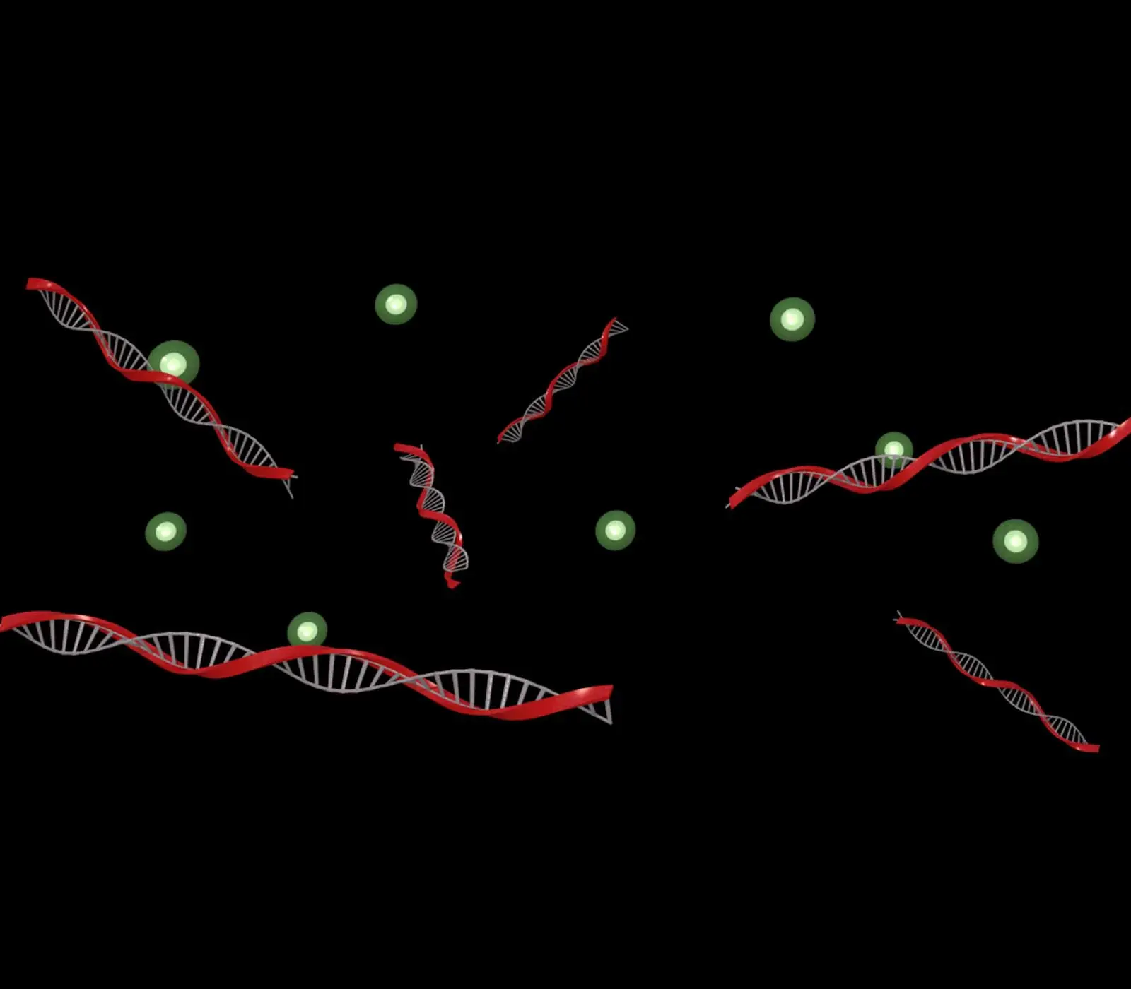
Labeling
Label your DNA or RNA probes and oligonucleotides using one-step direct methods, thiol-based indirect labeling, or specific 5′ and 3′ end tagging techniques.
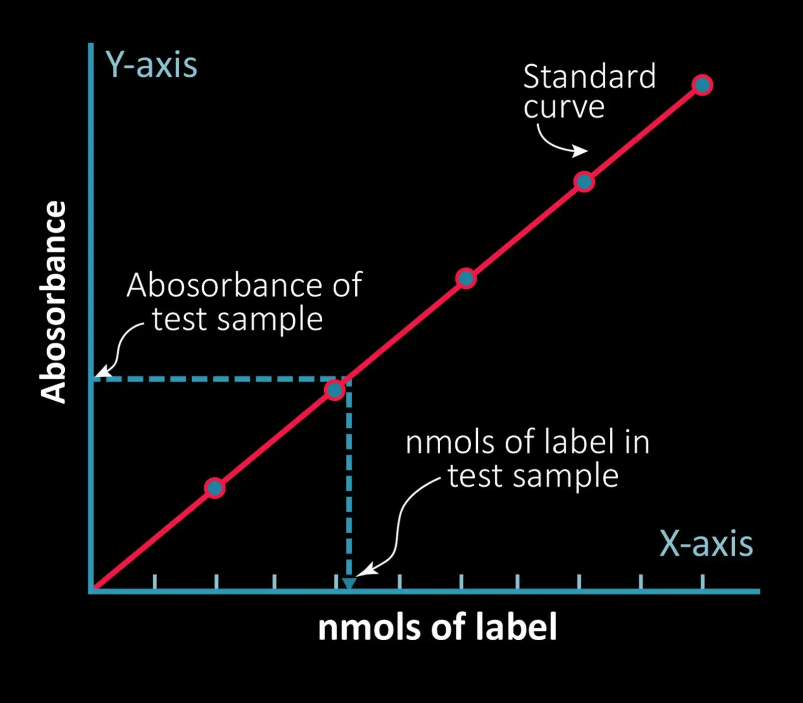
Characterization
Confirm probe customization and label incorporation. For example, use the QuantTag Biotin Quantitation kit to determine the number of biotin molecules incorporated in each strand of nucleic acid.
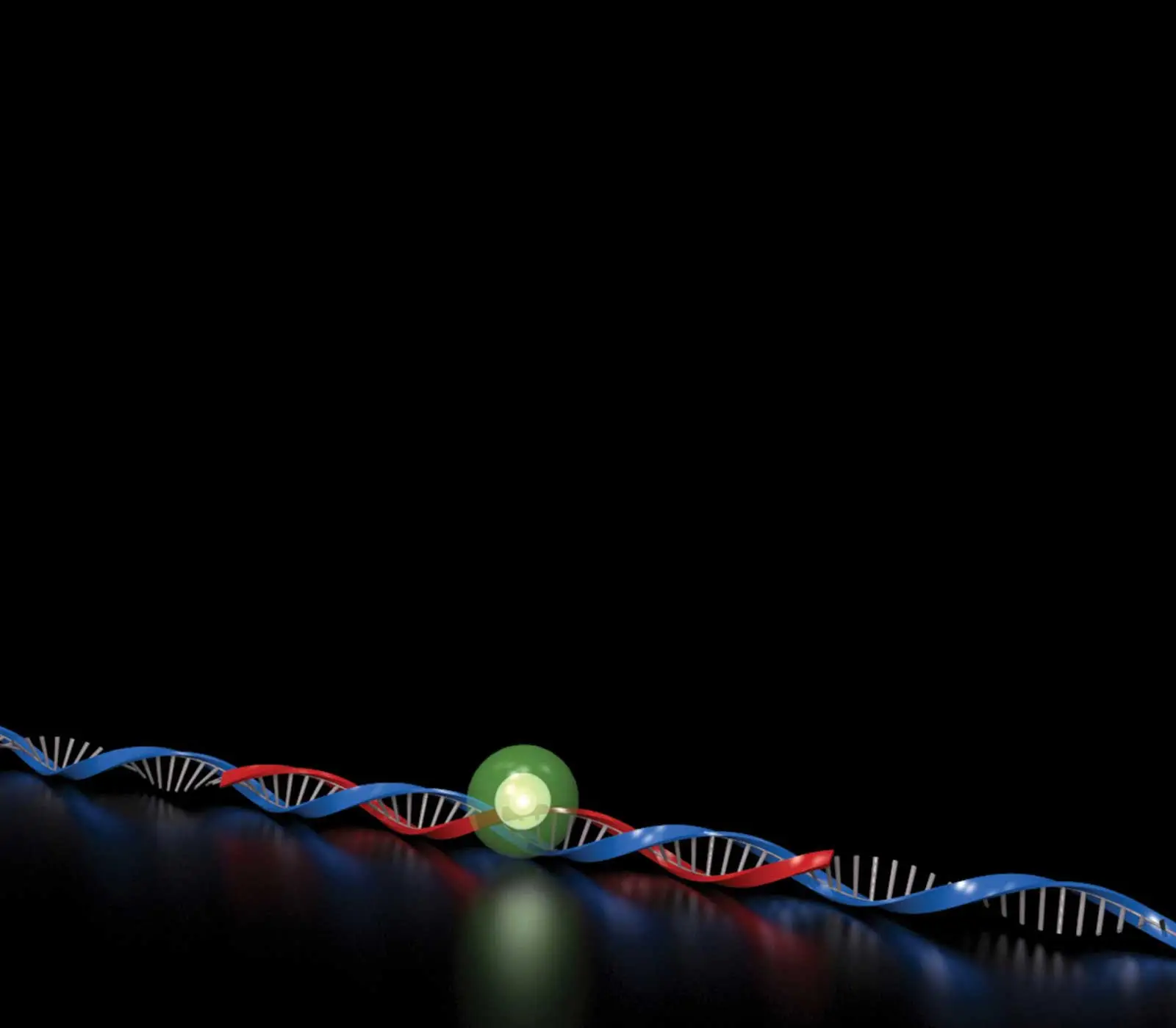
Hybridization
Optimize the probe concentration and buffer conditions to ensure effective annealing to complementary sequences.
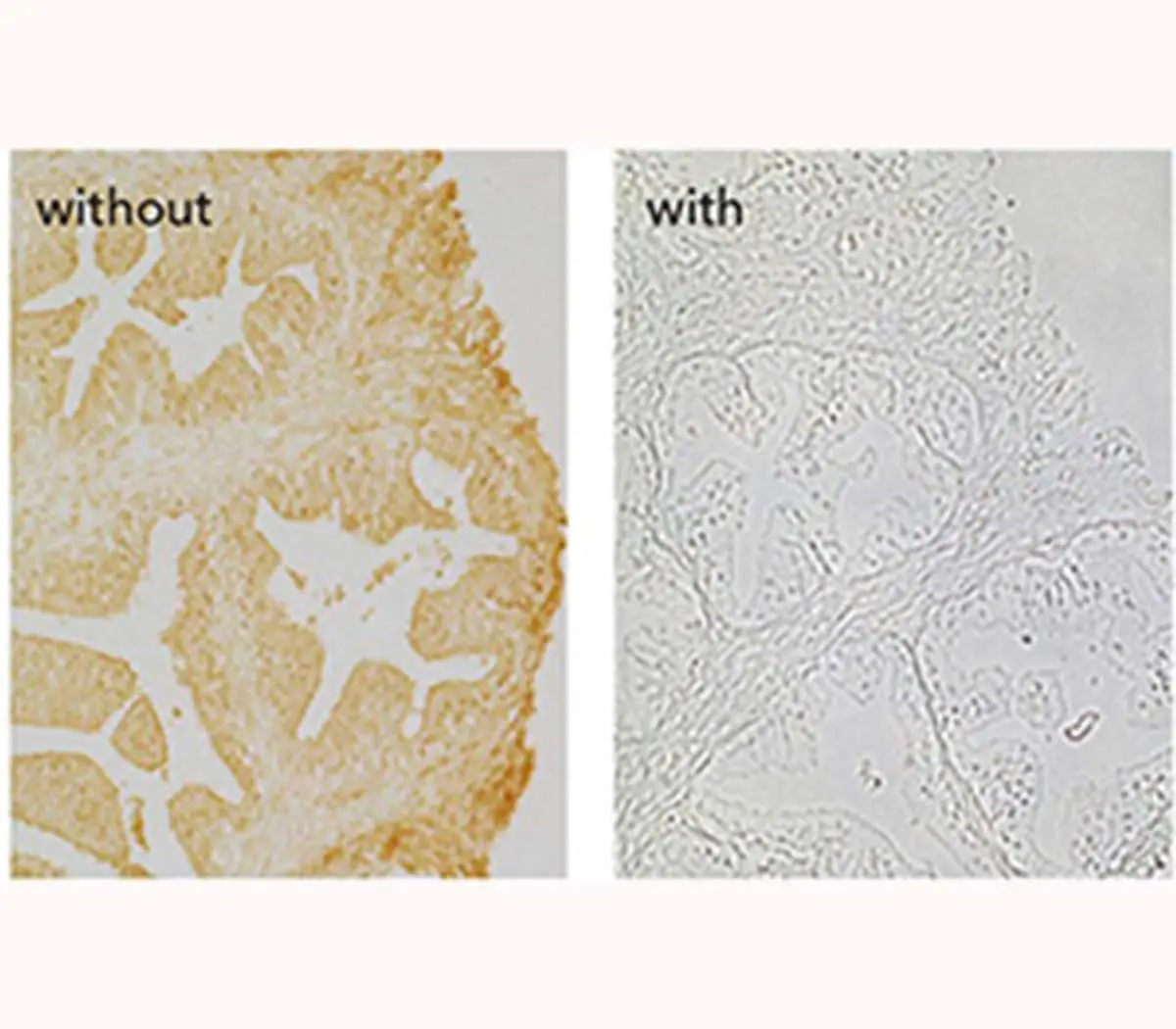
Blocking
Reduce or eliminate unwanted background (non-specific) staining on tissue sections and cell preparations using a blocking solution. Non-specific staining can result from endogenous enzyme or tissue elements, including endogenous enzyme activity, presence of Fc receptors, or interactions of detection reagents with tissue or cell proteins and other macromolecules. Choose a blocking solution based on the results of negative control sections.
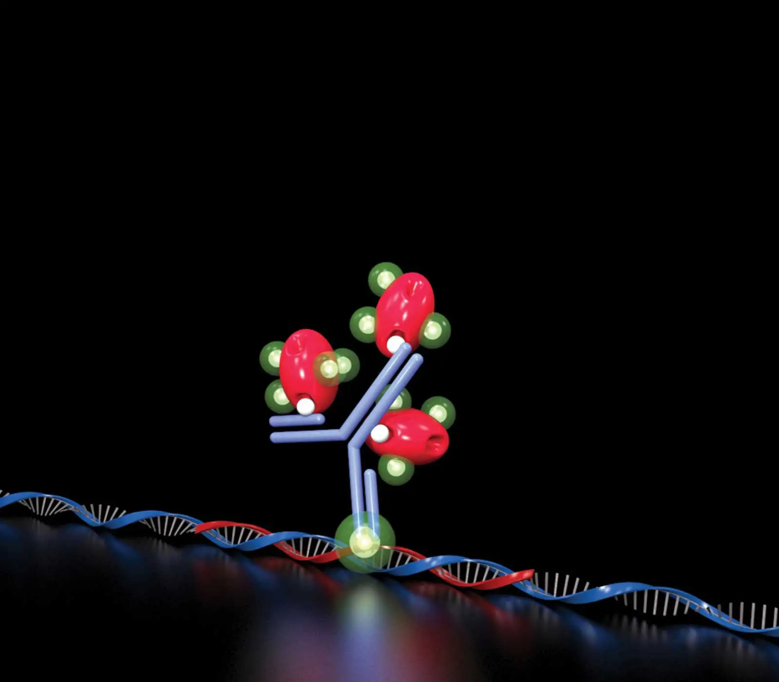
Detection
Use fluorescent or enzyme-conjugated reagents that meet your sensitivity requirements and visualization preferences.
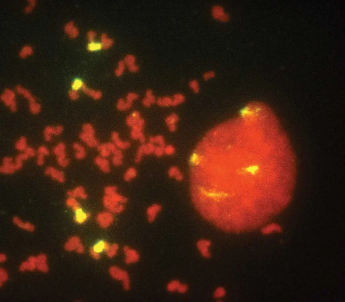
Counterstain and Mount
Retain and preserve the specific target signal for short-term storage or longer term archiving.

