
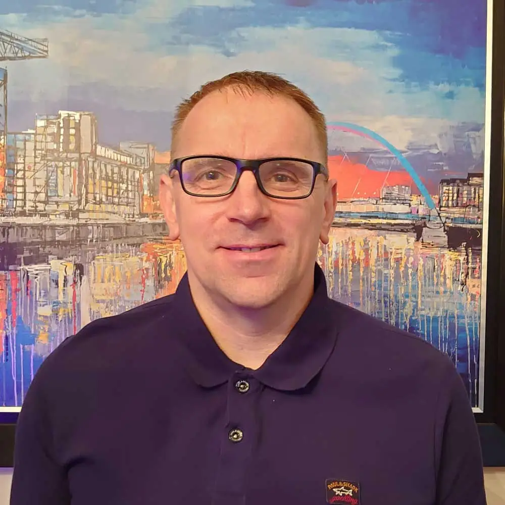
Not everyone had a talent for science as a child. Colin Nixon failed biology at school. He was described by most of his teachers as “academically challenged.” They likely would be surprised to see how Colin serendipitously “fell into” the sciences through a government youth training scheme that indicated he had an aptitude for the science, and he has since made a career of it. Today you can find Colin managing the Histology core lab at the Beatson Institute for Cancer Research in Scotland. Fourteen years ago, they approached him (he jokingly implies that it was a case of mistaken identity) to come aboard to set up a histology lab from scratch. Since then, he has been cultivating this facility into an essential core lab that provides service for cancer researchers.
“I’m a service provider for cancer researchers who provide me with bits of tissue. I will facilitate the processing of the tissue in a number of ways and give it back to them for analysis… I’m very fortunate that our institute…works across the board in all types of cancer…it makes it interesting that we don’t just specialize in just one kind of cancer.”
Colin was not the best student and dropped out of school with no real qualifications. However, he was pushed to find employment, and he ended up obtaining an ordinary national certificate in chemistry and applying for a histology job in a hospital. Despite it being a somewhat random choice of a job in a field that he initially “did not know or understand,” Colin was surprised to find that he was good at gross pathology and dissection and, what’s more, he enjoyed it. This first position led to an opportunity to progress as he landed a position at Glasgow University Veterinary School histology lab that helped support his continuing education all the way through to obtaining an MSc in Biomedical Science. To his surprise, he found that he loved working in the sciences “from the start.” With no managerial experience prior to developing the core lab, Colin claims that he had to learn how “NOT to treat people” and how to bring“ the best out of them.” But knowing that most of his team members stay in the lab for five to ten years, he must be doing something right.
“My only job has been in pathology since 1992. When I moved to Glasgow University Veterinary School in 1993, I diversified from diagnostic pathology of pets, cattle, and even the occasional tiger. From the recognition I got there, I was able to expand my work into animal research and to cancer research within different species. Mostly I work now in murine pathology or histology, but I also work in human cancer research in collaboration with local hospitals, have figured out how to work with Drosophila, and work on cellular material as well.”
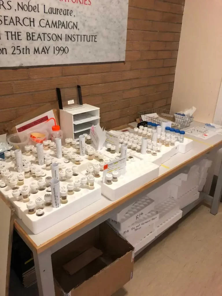
The Histology core facility sees a myriad of tissues and cancer types, with a focus on breast, liver, pancreatic, and colorectal tissue. The facility serves approximately 160 researchers within the Beatson Institute, Glasgow University, and around the world. Their services are always in demand, so much so that there usually is a backlog of work. To help keep research going throughout the pandemic, Colin fought to keep the lab open with seven-day rotations to safely accommodate his team with adequate social distancing. He was able to change that back to a five-day, nine-to-five workweek with staffing adjustments. Despite not being directly involved in research projects, he has been an author on 17 publications just since the beginning of the pandemic. In this contributing role, he adds his expertise to the development of content in the materials, methods, and protocols sections—sometimes developing new techniques. For Colin, writing for publications has turned out to be an unexpected source of enjoyment, and he has found that the researchers very much appreciate it.
Although Colin thought that by now histology would be on the decline, he certainly had good timing when he entered the field.
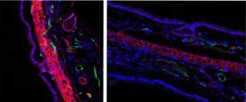
The advances in instrumentation and staining technology have been a bit of a shock to him. He envies the galaxy of colors possible in multispectral analysis seen at some of the meetings he attends and sometimes wishes that his lab could spend more time on certain techniques that help push the boundaries further. He sees future potential for his core facility clients, though, especially when the time-to-results shortens enough to become a part of the service provider menu. With cancer research remaining as vital as ever to advancing public health, Colin sees the promise of what old-school histological techniques and innovative technology can bring to light and looks forward to the next part of that journey.
“I’ve been doing my current job for 14 years and it’s passed in a blink of an eye. I’ve been on the campus of Glasgow University for nearly 27 years. It was because my current job used to send samples to my previous lab that they became aware of me. I think they thought I was someone else, and then they approached me to see if I’d like to set up a lab… People seem to keep me around for a while. I’m not sure if they’re scared to tell me to leave… I’m happy to stay where I am… I built this lab up from nothing with help from some great colleagues to a lab that gets a lot of praise. Any praise should go to the staff in the lab who put up with my demands and do the work otherwise we wouldn’t get the recognition we do.”





Stay in the Loop. Join Our Online Community
Products
Ordering
About Us
Application
Resources
©Vector Laboratories, Inc. 2025 All Rights Reserved.
To provide the best experiences, we use technologies like cookies to store and/or access device information. Consenting to these technologies will allow us to process data such as browsing behavior or unique IDs on this site. Not consenting or withdrawing consent, may adversely affect certain features and functions. Privacy Statement