Slide Preparation
Showing all 3 results
| Product | Image | Applications | Counterstain | Price / SKU | Select |
|---|---|---|---|---|---|
|
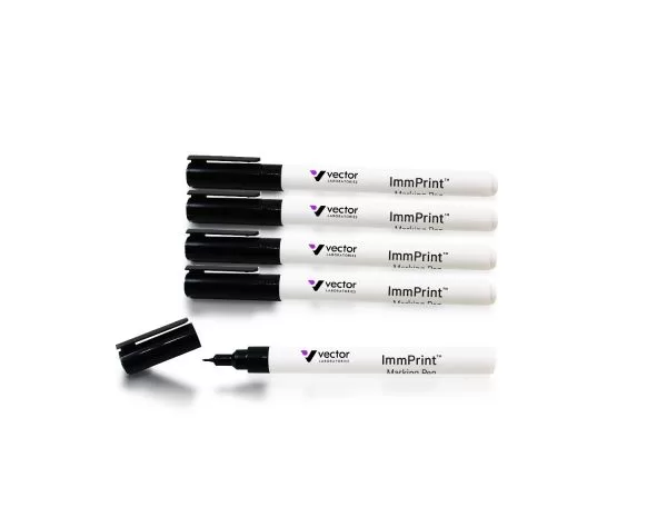
Immunohistochemistry / Immunocytochemistry, Immunofluorescence, In situ hybridization
|
None
|
$104.00
SKU: H-6100
Quantity:
|
|
|
|
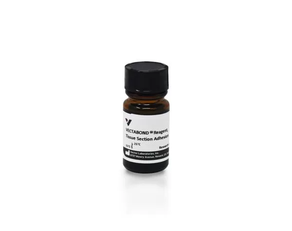
Immunohistochemistry / Immunocytochemistry, Immunofluorescence, In situ hybridization
|
None
|
$153.00
SKU: SP-1800-7
Quantity:
|
|
|
|
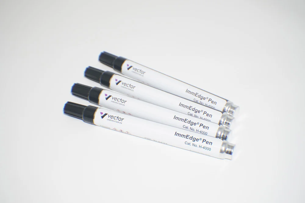
Immunohistochemistry / Immunocytochemistry, Immunofluorescence, In situ hybridization
|
None
|
$189.00
SKU: H-4000
Quantity:
|
|
No selected.
$0.00
