


| Unit Size | 15 ml, 50 ml |
|---|---|
| Applications | Immunohistochemistry / Immunocytochemistry, In situ hybridization, ELISAs |
| Technology | Micropolymer Reagents |
| Detection Enzyme(s) | Peroxidase |
| Conjugate | Micropolymer HRP |
| Target Species | Mouse |
| Format | Ready-to-Use |
The ImmPRESS Excel kits are a two-step peroxidase polymer based detection system that include an unconjugated amplifier antibody. This intermediate amplifier antibody increases sensitivity of the assay at least three- to four-fold over that of a one-step polymer based system, by facilitating the introduction of more peroxidase enzyme at the site of specific antigen localization. This increase in sensitivity would be advantageous in instances of weak antigen expression, and to further dilute out an expensive primary antibody. The ImmPRESS Excel Amplifier kits are presented as a complete kit format that include enzyme quench, blocking serum and ImmPACT® DAB EqV peroxidase substrate, in addition to the amplifier antibody and defined ImmPRESS polymer secondary antibody. However, as the two published references below indicate, this format is still modular and allows for the substitution of different detection reagents. These references describe using ImmPACT NovaRED™ peroxidase substrate in combination with the ImmPRESS Excel Amplifier kits. Oncogenesis (2017) 6, e293; doi:10.1038/oncsis.2016.82 Tan, E.M.S., et al (2017) Front. Med. 4:162 (doi: 10.3389/fmed.2017.00162)
Yes, the same ImmPRESS peroxidase secondary detection reagent can be used for double staining. There are a few considerations. First, we would suggest optimizing each single stain first on different tissue sections. Once the conditions for each stain have been established, the single assays should be run sequentially, from primary antibody though substrate color development, to achieve double staining on the same section. Secondly, different peroxidase substrates will have to be used to provide adequate color contrast and reliable target antigen localization. This double staining approach is most reproducible when detecting antigens expressed in different cell types on the same section, or different cell compartments of the same cell type. Please see our multiple antigen labeling guide for additional considerations:
Yes, we conducted studies on the compatibility of the ImmPRESS HRP polymer reagents with three commercially available autostainers (Agilent/Dako Autostainer Plus, Leica Bond Rx, and Ventana Discovery Ultra). We showed that our reagents are suitable for IHC detection on each of these platforms. The ImmPRESS polymer reagents and enzyme substrates generated equivalent IHC staining results compared to reagents from the instrument manufacturer. Some modifications in the protocol were performed to optimize the signal-to-noise ratio, such as a shorter incubation time for the ImmPRESS polymer and increase in the number of buffer washes following polymer incubation. Please see the following application note for more details about each automated platform: ImmPRESS App Note
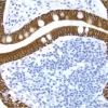
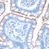
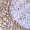
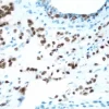
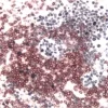
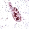
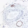
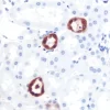
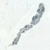
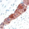
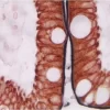
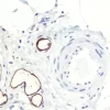
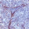
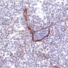
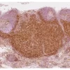
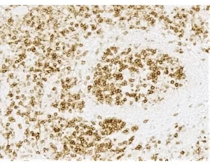
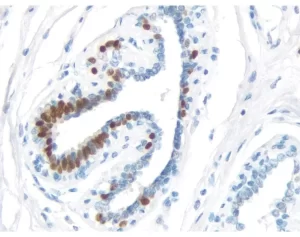
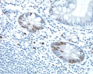
The peroxidase micropolymers of the ImmPRESS HRP polymer reagent limit steric interference and provide enhanced accessibility to the target, avoiding the disadvantages of other polymer systems that use large dextrans or other macromolecules as backbones. The result is crisp, strong staining of antibody targets, especially nuclear and membrane antigens (such as Ki67, estrogen receptor, bcl-2, CD3, CD8 and CD10) and greater sensitivity than other polymer systems.
The staining procedure is simple as shown in the diagram below. Following a blocking step with the diluted normal horse serum, sections are incubated with primary antibody. After a brief wash, the appropriate ImmPRESS Reagent is added to the sections and incubated for 30 minutes. Sections are again rinsed and the slides are developed with the peroxidase substrate of choice.

Consider Species Cross-Reactivity
When choosing the optimal detection system for your application, it is important to consider not only the species of the primary antibody but also the species of the tissue. If the species of the primary antibody and the species of the tissue are closely related (e.g. rat and mouse), the secondary antibody may bind to endogenous IgG in the tissue section leading to background. The following options minimize background staining in these instances:
Product review on Biocompare.com.


 Powered by Bioz © 2023