Vector Laboratories is closed for the President’s Day on Monday, February 19th. We will be back in the office on Tuesday, February 20th.
We will respond to emails upon our return. Have a wonderful day.
Menu
Vector Laboratories is closed for the President’s Day on Monday, February 19th. We will be back in the office on Tuesday, February 20th.
We will respond to emails upon our return. Have a wonderful day.

Both lectins and antibodies are powerful binding proteins used across many applications. Since their discovery in the late 19th century, these molecules have been widely studied, classified, and utilized in the advancement of research, health, and the sciences. The following article highlights key similarities and differences between lectins and antibodies and the types of analyses where they might be applied.
Lectins are molecules that bind to carbohydrates, also called glycans. Each has a unique affinity towards a particular carbohydrate structure. For example, Maackia Amurensis Lectin I (MAL I) has preferred sugar specificity towards Galβ4GlcNAc. The wide variety of lectins allows for complex evaluations on the glycome level.
Antibodies are made in an immune response to have direct affinity towards a specific antigen. An antigen is a molecule that can induce an immune response and is bound to an antibody. Antigens can be proteins, small peptides, or even other antibodies.
Lectins are found in various organisms including algae, fungi, bacteria, animals, and plants. Their origin determines the binding properties and therefore their function. For example, bacterial lectins bind to surface glycans at the terminal sugar or internal oligosaccharide chains. These lectins are crucial to bacteria for recognizing glycan sequences on the host cell for both pathogenic and symbiosis purposes.
Those derived from plants are the best-studied and most utilized because of their abundance in nature and ease of extraction. Check out our blog post, “Everything We Know About Lectin Structure, Classification, and Function,” for a more in-depth explanation of each lectin type.
Antibodies are naturally produced in B cells of animal immune systems. They are released into the bloodstream and lymph system, allowing for extraction via the serum. Those purified from the serum of an immunized animal are called polyclonal and will have a mixture of epitope targets of the same antigen.
Alternatively, monoclonal antibodies can be produced in a laboratory. In this process, individual B cells are isolated from an immunized animal, cloned, and grown to produce homogenous antibodies. These monoclonal antibodies not only target the same antigen, but a single epitope, aka binding site, on the antigen.
Lectins come in a wide range of shapes and sizes, giving a range of carbohydrate-binding sites. They can be classified structurally based on the number of binding sites.
Merolectins have a single carbohydrate binding domain. Hololectins contain 2 or more identical domains. Superlectins possess 2 non-identical domains. Finally, chimerolectins have both an enzymatic domain and a carbohydrate-binding domain.
In addition to domains, lectins are commonly classified according to the monosaccharides for which they have the highest affinity: mannose, galactose or N-acetylgalactosamine, N-acetylglucosamine, N-acetylneuraminic acid, or fucose.
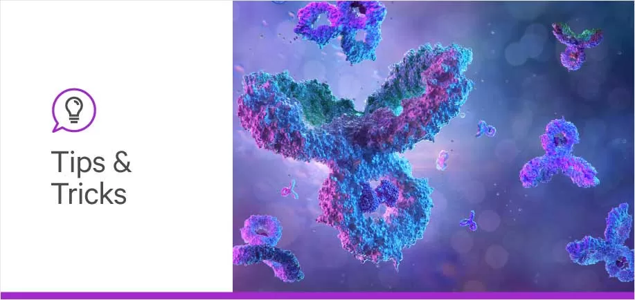
The standard antibody is a Y-shaped protein composed of 2 heavy chains and 2 light chains. The Fab fragment contains both light chains and portions of the heavy chains. This is the region that binds to the antigen and therefore has high variability. The Fc region is the tail composed of only the heavy chains portion. This is a highly conserved region allowing antibodies to interact with cell surface receptors.
The type of heavy chain found in antibodies distinguishes them into 5 classes: IgG, IgM, IgA, IgE, and IgD. Each class has a unique structure allowing for specific biological properties, locations, and antigenicity.
Lectins are used in glycobiology studies to profile, characterize, and capture complex glycans in biological systems. They can also be used as biomarkers for various diseases and conditions, as their expression levels can vary in response to physiological changes. Antibodies are used in various methods of biological research for identifying and characterizing a specific antigen.
Antibodies and lectins can be used in a similar fashion in a variety of techniques including, but not limited to, immunohistochemistry, immunofluorescence, ELISA, flow cytometry, immunoprecipitation, and affinity matrix.
While they are both detection agents, they will differ in the applicable assay due to lectins binding to glycans and antibodies’ affinity for antigens. For example, many diseases cause a change in the glycosylation of cells and the glycoproteins present on the cell membrane.
To study the glycosylation in normal, diseased, and treated cells, it would be more valuable to employ a lectin assay, which is specific to glycans, over an antibody. Additionally, when branching into the unknown territory of the effects of new diseases or treatments, lectin screenings can be performed to determine how disease/treatment alters the glycan profile.
Alternatively, antibodies would be a more beneficial tool when studying a specific protein, especially if the exact glycosylation of the protein is unknown. Additionally, the use of antibodies would be necessary over lectins in a double detection assay where both targets have the same glycan expression. Lectins would have trouble distinguishing the two, but antibodies could be more specifically directed at each target.
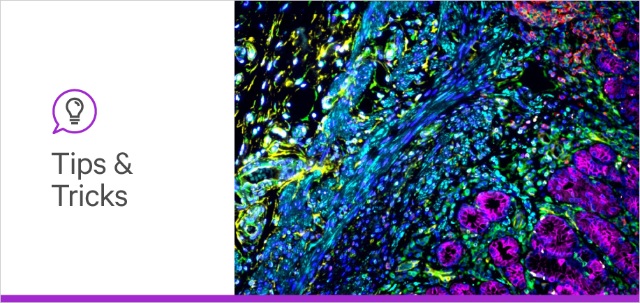
Experiments can even harness the power of antibodies and lectins together. For example, Lycopersicon Esculentum (Tomato) Lectin (LTL) is an effective marker of blood vessels in rodents. Ki67 is a protein marker that indicates cell proliferation and can be directly identified by an anti-Ki67 antibody. A study could evaluate new blood vessel development after an infarction. The antibody will mark Ki67 to highlight proliferating cells while the LTL will mark all existing and new blood vessels.
We hope this blog post helped you learn which detection protein is the right choice for your workflow. For more tips and tricks to push your research forward, stay tuned to the blog


Stay in the Loop. Join Our Online Community
Together we breakthroughTM

©Vector Laboratories, Inc. 2024 All Rights Reserved.