
Vector Laboratories is closed for the President’s Day on Monday, February 19th. We will be back in the office on Tuesday, February 20th.
We will respond to emails upon our return. Have a wonderful day.
Menu
Vector Laboratories is closed for the President’s Day on Monday, February 19th. We will be back in the office on Tuesday, February 20th.
We will respond to emails upon our return. Have a wonderful day.
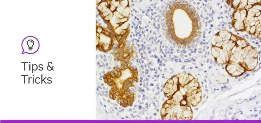
Fixation is critical for later processing and analysis of your tissue sample, but, in some cases, it can create barriers blocking the information you need most. There’s nothing worse than spending days on an immunostaining experiment and looking at your slide under the microscope, only to be faced with weak or no staining.
Instead of wallowing in despair at the results, the culprit might be the fixation method itself. If you are working with paraffin-embedded tissue sections, it is likely that your epitope of interest is masked and therefore unavailable for antibody binding. If this sounds like your situation, then fear not! Heat-Induced Epitope Retrieval (HIER) may be the solution to your problems. HIER is a pre-staining treatment used to restore secondary and tertiary structure for enhanced antibody binding. In this way, epitopes become “unmasked” and available for your antibody to bind, increasing detection. You will need to optimize the protocol for your tissue depending on the antibody, epitope, heating apparatus, fixation method, and tissue type.
In this article, we have some tips to get you up and running with antigen retrieval to enhance your staining and alleviate your stress.
Formalin tissue fixation can lead to protein cross-linking or conformational changes by introducing methylene bridges, which may mask the epitope you are trying to detect and reduce primary antibody binding. Antigen retrieval reverses these processes, allowing the antibody to bind onto the antigen. If you have weak or absent staining, this can be an indicator that antigen retrieval may be required.
Antigen retrieval is a technique that unmasks an epitope, restoring the antibody’s ability to bind. It is used on tissues where antigens are cross-linked or otherwise impacted by the fixation process. Antigen retrieval is more likely to be needed when using monoclonal antibodies versus polyclonal antibodies, as polyclonal antibodies can bind to multiple epitopes whereas monoclonal antibodies only bind one. However, polyclonal antibody staining can often be improved with antigen retrieval. Either way, antigen retrieval can help you unlock the secrets hidden in your tissue with consistent staining results.
Now that you’ve taken a deep breath, accepted your fate, and decided to try antigen retrieval, there are three main considerations for protocol optimization, dependent on equipment and buffers: pH, temperature, and incubation time. Usage instructions from the primary antibody manufacturer should offer some guidelines on where to start.
It’s important to note that excessive heating temperature and duration can lead to tissue damage, so be sure to start low and slow, increasing your parameters incrementally — it’s all about finding the optimal middle ground to unmask your epitope without destroying your tissue!
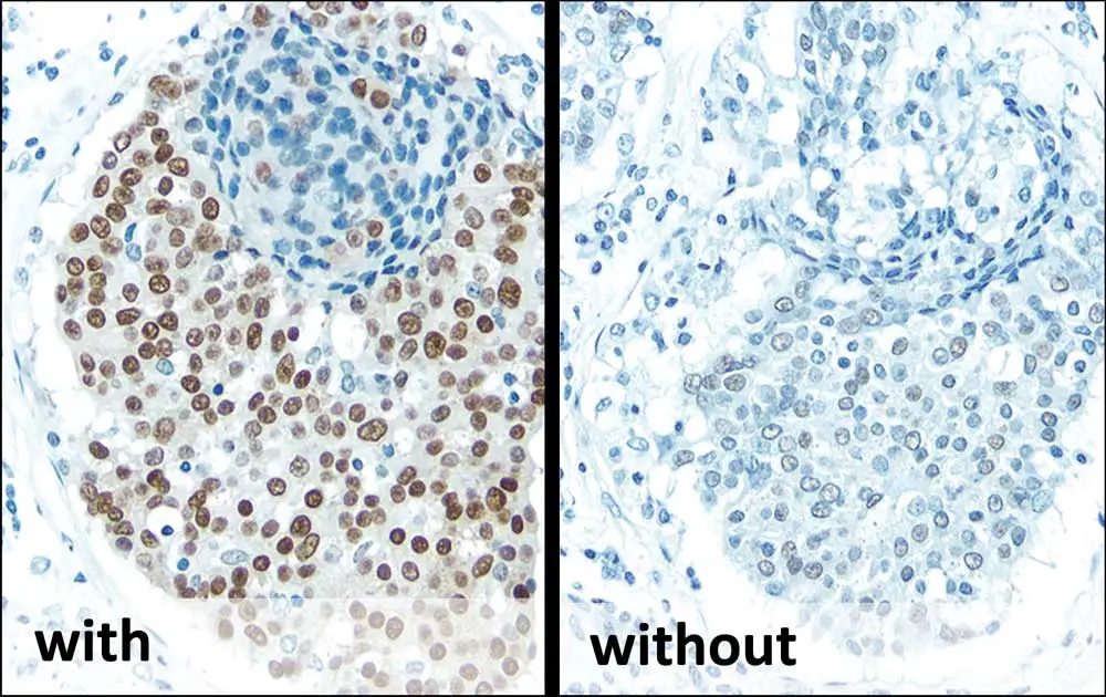
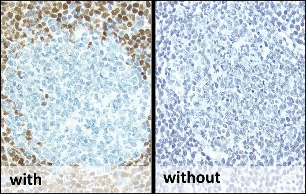
A good starting point is to refer to the primary antibody manufacturer’s product specification to confirm that the antibody has been tested and validated for your specimen species and application (immunohistochemistry versus western blot, for example). Next, we recommend performing some control experiments to verify that your detection system is working properly. If your detection system is not to blame, then you may need to re-optimize your antigen retrieval conditions or explore other fixation methods for your specimen.
Yes! To help you on your antigen retrieval journey, be sure to check out Vector Laboratories’ Citrate-Based (pH 6.0) and Tris-Based (pH 9.0) Antigen Unmasking Solutions, which are highly effective at unmasking epitopes from formalin-fixed, paraffin-embedded tissue selections when combined with HIER, which is a pre-staining treatment used to restore secondary and tertiary structure for enhanced antibody binding.
Be sure to check out our IHC Resource Guide or contact our Technical Support if you need additional help.


Stay in the Loop. Join Our Online Community
Together we breakthroughTM
©Vector Laboratories, Inc. 2024 All Rights Reserved.