Format Ready-to-Use | |
Unit Size 1 Kit | |
Storage Instructions (15–25° C) | |
Applications Immunohistochemistry, Immunofluorescence | |
Target Species Universal | |
Source Species Universal |


VectaPlex™ Antibody Removal Kit
Your path to simplified multiplexing starts with VectaPlex™. This kit is designed to remove antibodies and other non-covalently bound detection reagents from FFPE tissue sections in immunohistochemistry (IHC), immunofluorescence (IF) and tyramide signal amplification (TSA) workflows.
Benefits include:
- Removes antibodies and other non-covalently bound detection reagents from FFPE tissue sections
- Compatible with immunofluorescence-, fluorescent tyramide- and immunohistochemistry-based workflows
- Obtain more spatial biomarker information from each tissue section
- Stain with multiple primaries from the same species and a single secondary without cross reactivity
- Maintains tissue morphology through multiple cycles – even on delicate specimens like breast & skin
- Maintains tissue antigenicity through multiple cycles
- Primary antibody concentrations do not need to be optimized based on the order of detection in multiplex workflows
- Easy-to-use, room temperature application
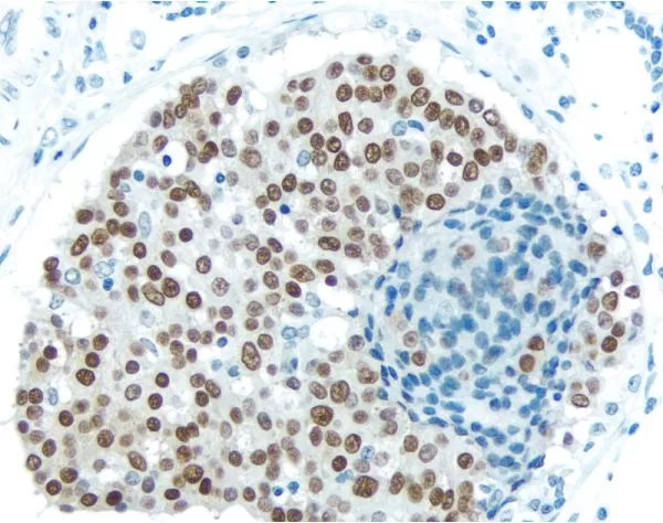
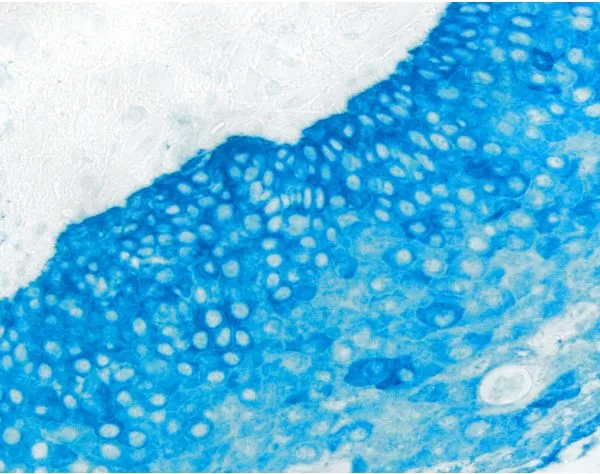
The first image above is a composite image generated from individual images taken during six rounds of immunofluorescence staining using VectaPlex. FFPE stomach; six mouse primary antibodies – CD20 (red), CD34 (yellow), Desmin (cyan), AE1/AE3 (magenta), CD3 (gray), Vimentin (green) – detected with DyLight™ 488 horse anti-mouse IgG. Blue indicates DAPI stain. Vector® TrueView® Autofluorescence Quencher Kit applied to suppress background fluorescence.
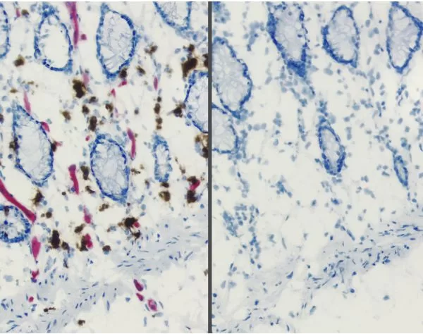
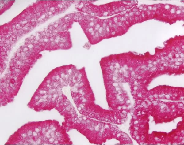
The second image above is a 4-plex IHC image taken of esophagus tissue stained for CD68 (brown) using ImmPACT DAB, CD20 (red) using ImmPACT Vector Red, CD34 (blue) using Vector Blue, and Desmin (grey) using ImmPACT SG. Each round of staining was performed using normal IHC protocols with VectaPlex treatment between each round of staining.
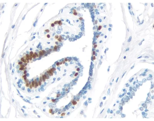
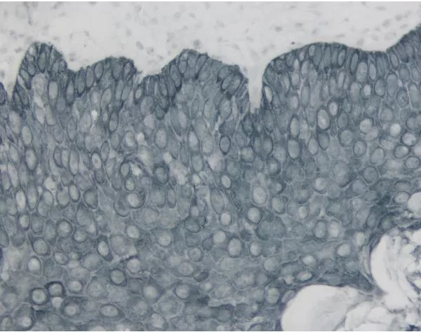
The third image above was captured after three rounds of tyramide staining. VectaPlex was used after each round. FFPE tonsil is shown, following application of 3 mouse primary antibodies – CD3 (red), CD68 (green) and AE1/AE3 (purple) – detected using Opal 3-plex TSA Manual Detection Kit. Blue indicates DAPI stain.
$312.00
| SKU | Unit Size | Price |
|---|---|---|
Select a unit size:
How do I request a quote or bulk pricing?
Technical Information This ready-to-use kit is designed to remove antibodies and other non-covalently bound detection reagents from formalin-fixed paraffin-embedded (FFPE) tissue sections in immunofluorescence or tyramide signal amplification staining workflows. VectaPlex simplifies sample processing for manual workflows, accelerates multiplex assay optimization, preserves precious sample, and maintains tissue integrity and morphology through multiple cycles of antibody binding and removal. VectaPlex IHC Workflow: After staining with one of the approved substrates, VectaPlex is used to remove detection reagents in preparation for another cycle of staining. The substrate is not removed or impacted by VectaPlex. A single image is collected after all cycles are complete.
|
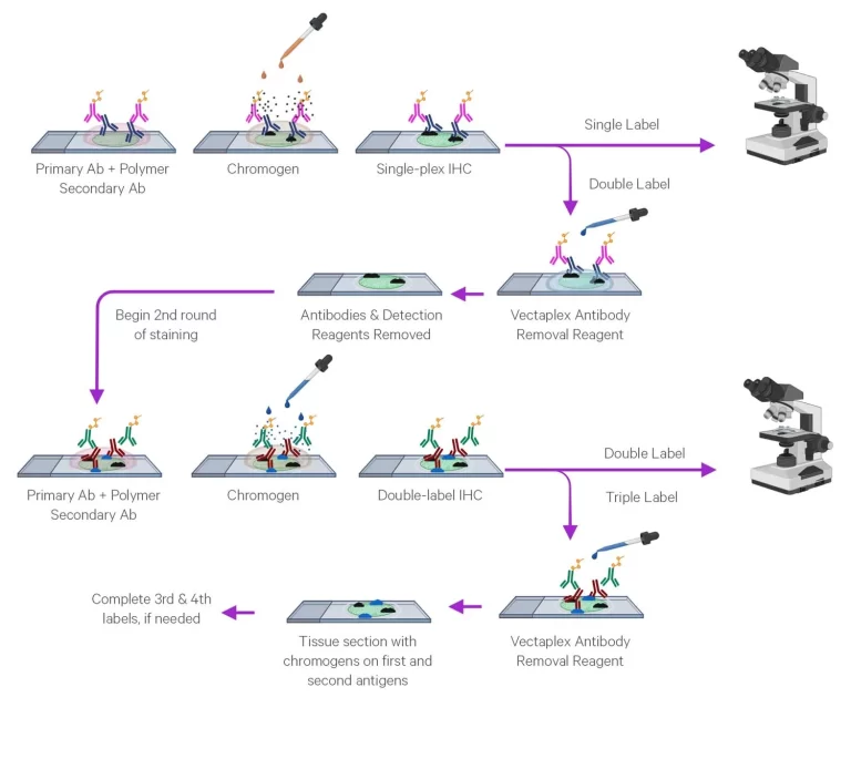 VectaPlex IF Workflow: After IF staining and imaging the sample, VectaPlex is used to remove detection reagents in preparation for another cycle of staining. After all images have been collected, a composite image is formed from individual images.
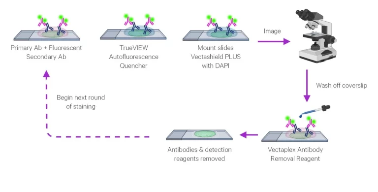 VectaPlex TSA Workflow: After TSA staining and imaging the sample, VectaPlex is used to remove detection reagents in preparation for another cycle of staining. The tyramide sigal is not removed by VectaPlex. A single image is collected after all cycles are complete.
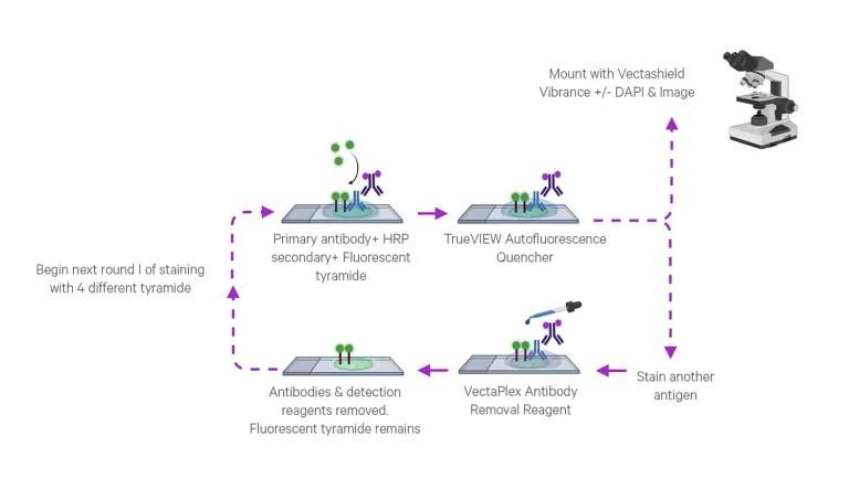
Product FAQs
Can other substrates be used with VectaPlex?
Perhaps. We have not tested substrates from other vendors but they may be compatible. To test this, simply stain two sections with the substrate in question and treat one of them with VectaPlex. If the staining intensity is unchanged, the substrate should be compatible with VectaPlex.
Can I use VectaPlex on frozen sections?
No. During development, VectaPlex was optimized for FFPE tissues, where the power to remove antibodies at room temperature was balanced with the need to preserve tissue morphology. On frozen tissues, VectaPlex treatment can lead to tissue damage and loss.
I am using TrueView to quench autofluorescence; will TrueVIEW be stripped by VectaPlex reagents?
We used TrueView in our VectaPlex IF workflow to suppress autofluorescence. Application of VectaPlex removes some, but not all, of the TrueView reagent. Since some TrueView remained after VectaPlex application, we did not add TrueView in every cycle, but only as needed to suppress autofluorescence.
I accidentally put my VectaPlex kit in the freezer. Can I still use it?
Yes. VectaPlex can withstand a freeze-thaw cycle and still performs as expected. However, the recommended storage temperature is at ambient (15–25 °C). Additionally, the kit will still perform properly if placed at 4 °C.
Is there a maximum number of times you can use VectaPlex on one section?
VectaPlex has been tested for up to 6 cycles with no adverse effects. We don’t expect there to be issues with increasing the number of cycles, but we suggest testing with controls for adequate comparisons. We recommend running experiments to confirm that the use of more cycles does not impact the antigenicity or tissue morphology.
Can VectaPlex be used on an autostainer?
Yes. We have validated this product for use on automated systems.
Does VectaPlex work for all species of antibodies?
VectaPlex has been tested with mouse and rabbit primaries, and with horse and goat secondaries. We expect it to work well with other species of antibodies as well.
Does VectaPlex work on different tissue types?
VectaPlex has been tested on and works well with various FFPE tissues, including FFPE breast and lung tissue which are very delicate.
Is VectaPlex compatible with all antibodies?
VectaPlex has been tested on more than 30 common antibodies and has worked on them without issue.
In an IF protocol, will the VectaPlex Antibody Removal Kit also strip the autofluorescence from the tissue?
VectaPlex does not reduce autofluorescence background. Vector’s TrueVIEW Autofluorescence Quenching Kits (SP-8400-15, SP-8500-15) effectively diminish unwanted autofluorescence from non-lipofuscin sources.
Is there a protocol for removal of coverslips between VectaPlex cycles when performing IF?
We recommend using VECTASHIELD® Plus Antifade Mounting Media with DAPI (H-2000), which is an aqueous non-hardening mounting medium. After each imaging cycle, the coverslip can be easily removed by incubating in PBS and the residual VECTASHIELD is removed with a PBS wash.