Blocking Action Autofluorescence | |
Format Concentrate | |
Unit Size 1 kit | |
Applications Immunofluorescence, In situ hybridization |


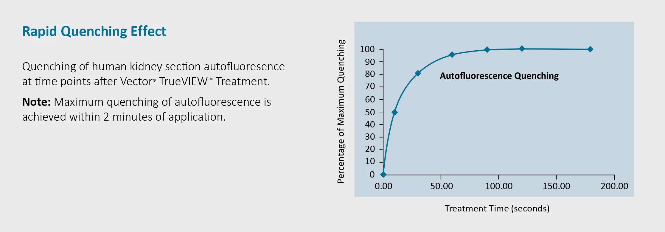
Vector TrueVIEW Autofluorescence Quenching Kit
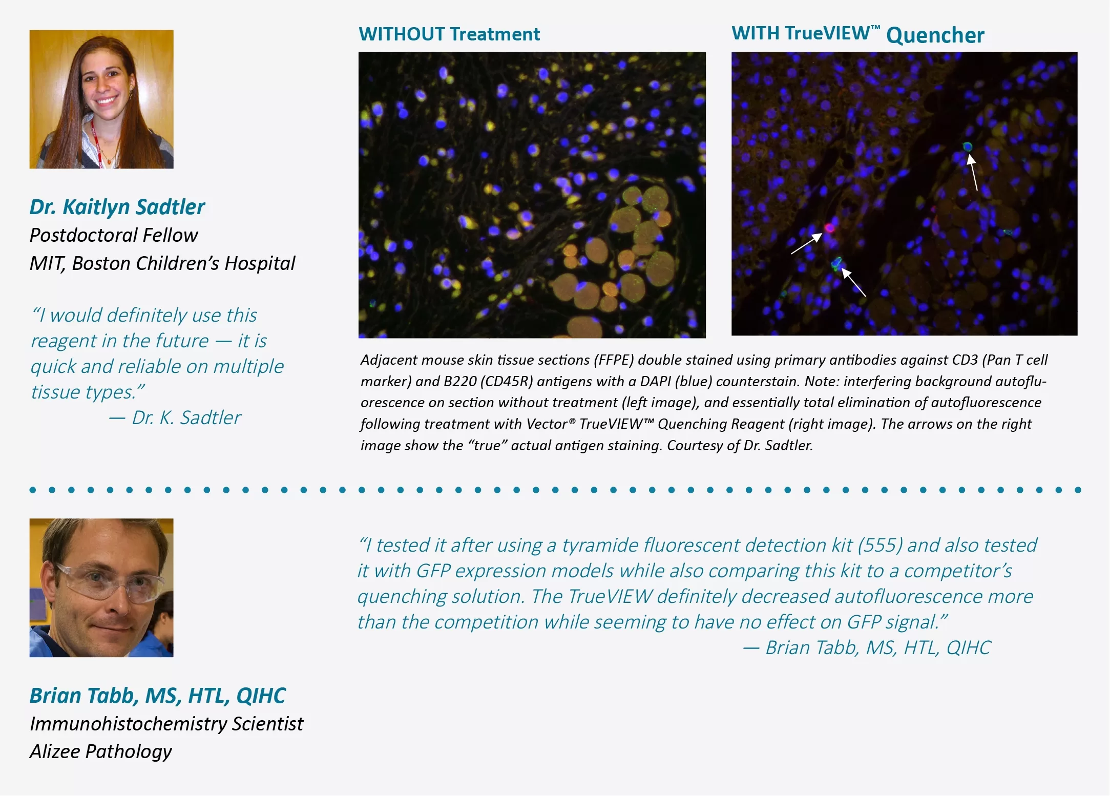
The TrueVIEW Autofluorescence Quenching Kit provides a novel way to diminish unwanted autofluorescence from non-lipofuscin sources and dramatically improve signal-to-noise ratio. TrueVIEW removes unwanted autofluorescence in tissue sections due to aldehyde fixation, the presence of red blood cells, and structural elements such as collagen and elastin. The quenching action of the kit reagents provides a clear, unambiguous, “true view” localization of the target antigen.
This kit offers you:
• Effectivity with problematic tissues such as kidney and spleen
• An easy-to-use, one-step method
• Compatibility with a wide selection of fluorophores
• Compatibility with standard epifluorescence and confocal laser microscopes
• Availability with DAPI counterstain and without.One kit is sufficient to treat approximately 100 to 150 tissue sections.
$173.00
| SKU | Unit Size | Price |
|---|---|---|
Select a unit size:
How do I request a quote or bulk pricing?
Kit Components What’s in the box?
|
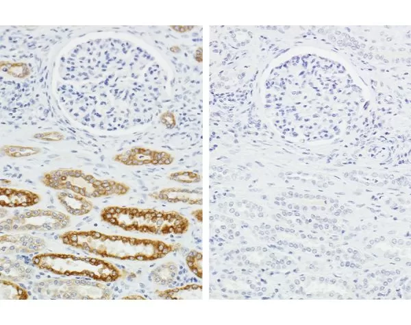
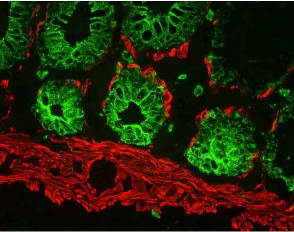
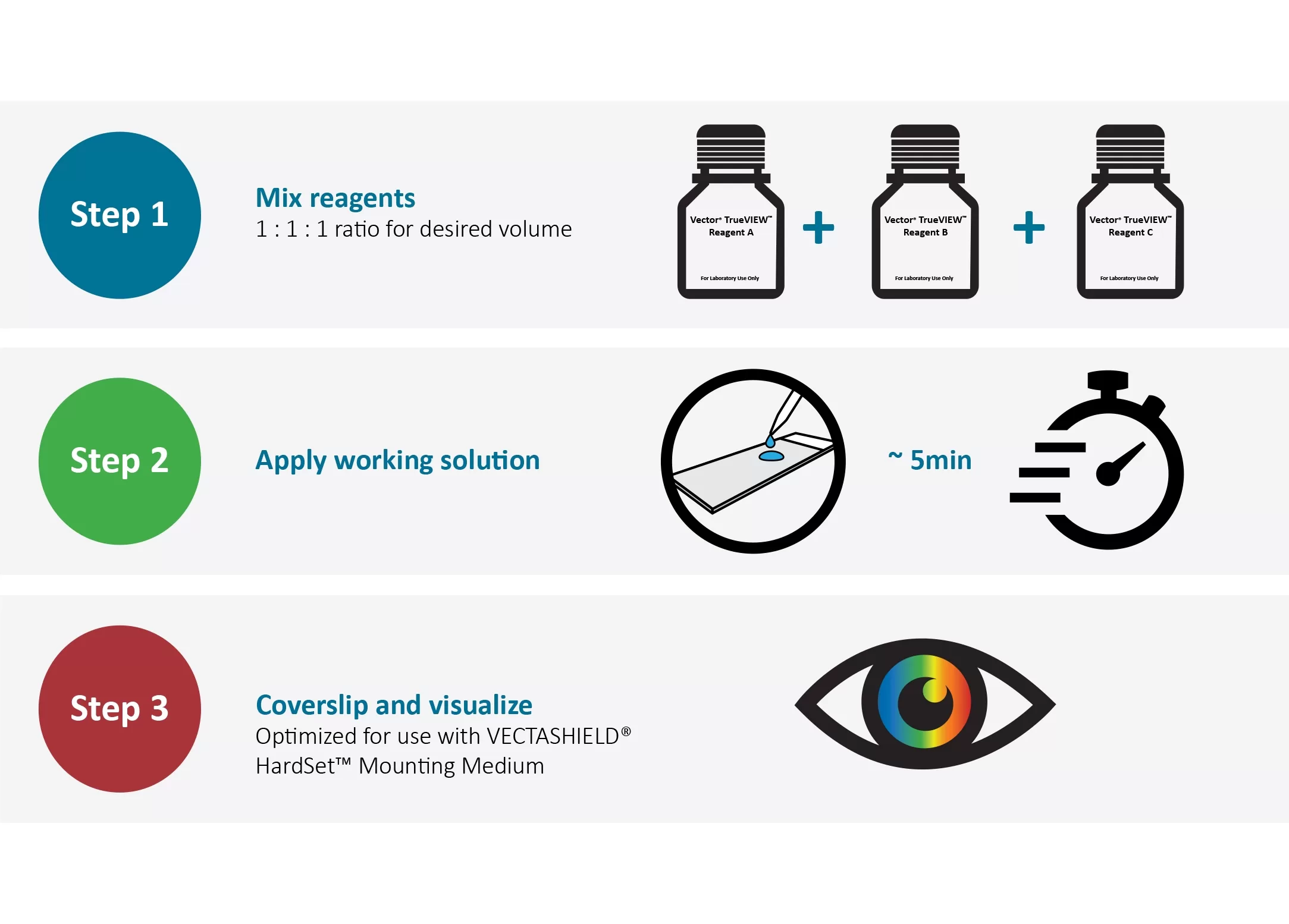
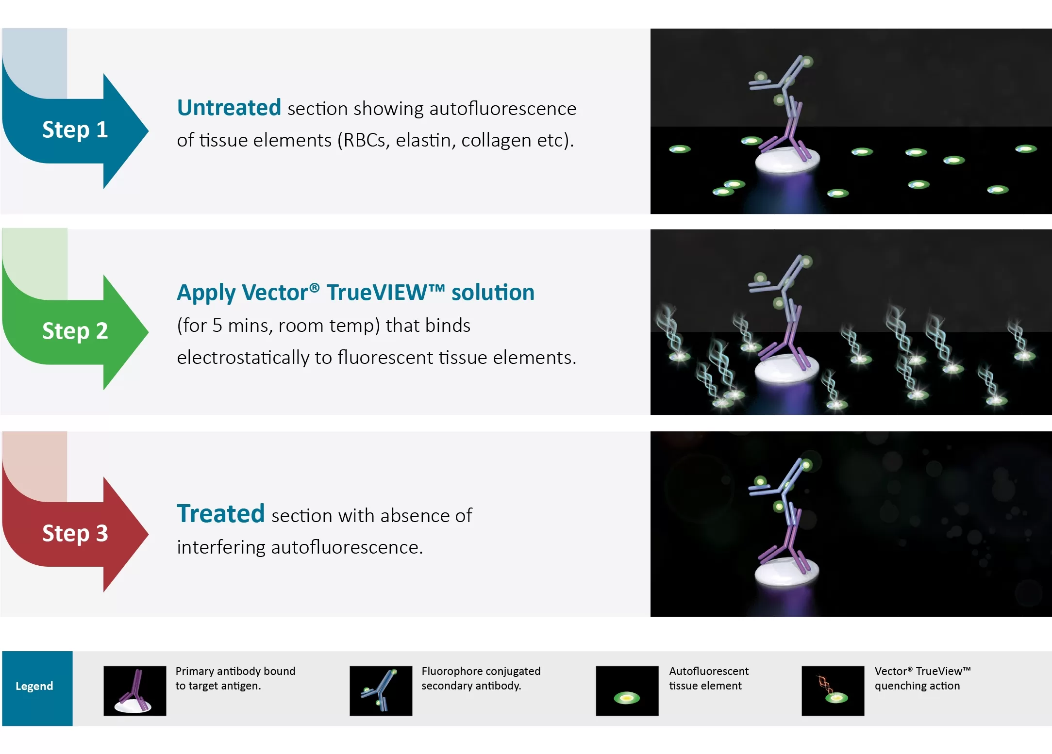
Technical Information Tissue autofluorescence often occurs with aldehyde fixation or from inherent native tissue components (collagen, elastin, and red blood cells). The extent and intensity of autofluorescence background frequently makes it difficult or impossible to distinguish specific signals in immunofluorescence applications. Most methods for reduction of tissue autofluorescence act primarily on lipofuscin granules, and are not broadly effective against the most common sources of autofluorescence targeted by TrueVIEW Quencher. Current methods for reducing autofluorescence primarily include “home brew” concoctions such as sodium borohydride and other ink-based products. These methods are essentially ineffective against aldehyde induced autofluorescence. In contrast, TrueVIEW reagent binds to hydrophilic compounds and effectively quenches endogenous autofluorescence. 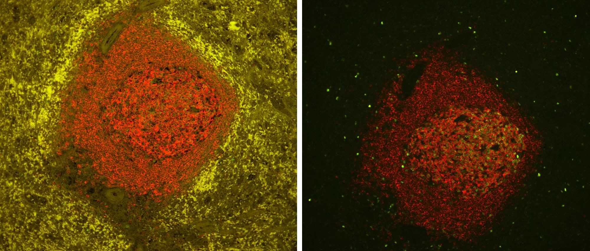 Image showing Spleen FFPE tissue stain, with & without TrueVIEW autofluorescence quencer. Antigen retrieved with Antigen Unmasking Solution. Spleen tissue stained with Mouse Anti-CD20 (red) and Rabbit Anti-Ki67 (green), followed by VectaFluor™ Duet Kit, and mounted in VECTASHIELD Vibrance Mounting Medium with DAPI. Left Image: No TrueVIEW Quencher treatment – 20x objective, red channel exposed 200ms, green channel 200ms. Note presence of interfering autofluorescence. Right Image: Cells treated with TrueVIEW Autofluorescence Quencher – 20x objective, red channel exposed 200ms, green channel exposed 800ms. Note absence of autofluorescence. Human tonsil (FFPE); Section stained for AE1/AE3 using fluorescein label (green). Treated with TrueVIEW and mounted with VECTASHIELD Vibrance Antifade Mounting Medium with DAPI (nuclei blue). 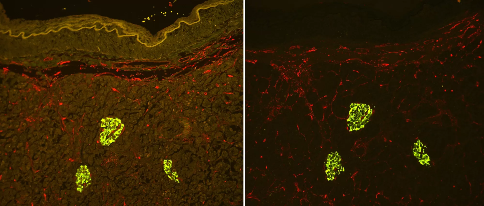 Image showing Pancreas FFPE tissue stain. Antigen retrieved with Antigen Unmasking Solution, stained with Guinea Pig x Insulin (green) and Mouse x END (red), followed by FL-Goat x Guinea Pig IgG + DyLight™ 594-Horse x Mouse IgG. Mounted in VECTASHIELD Vibrance Mounting Medium with DAPI. Left Image: No TrueVIEW Quencher used, 20x objective, red channel exposed 600ms, green channel 600ms. Right Image: TrueVIEW Quencher used with 20x objective, red channel exposed 600ms, green channel exposed 600ms. Easy To ApplyFollowing completion of the IF staining procedure:
Mode of ActionFollowing completion of the IF staining procedure:
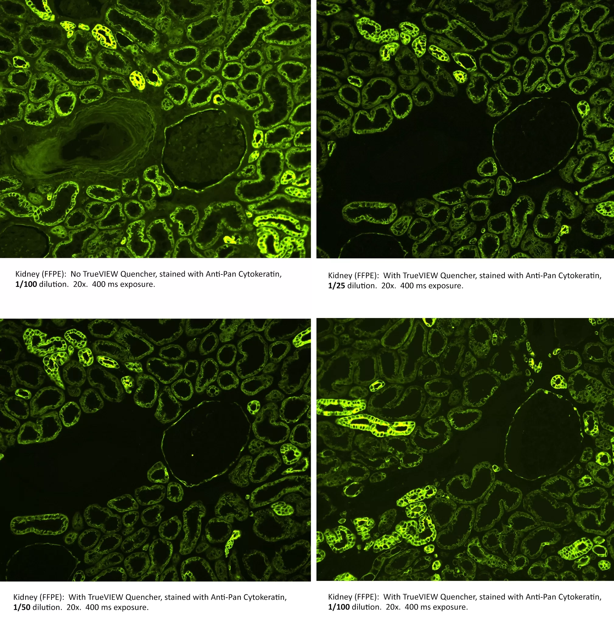
Signal to Noise OptimizationThe primary antibody and detection reagents should be optimized (titered) in conjunction with Vector TrueVIEW Quenching Kit to achieve maximum signal to noise.
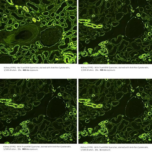
Optimization of ExposureIncreasing exposure times may be necessary to achieve optimal image acquisition for TrueVIEW Quencher treated slides.
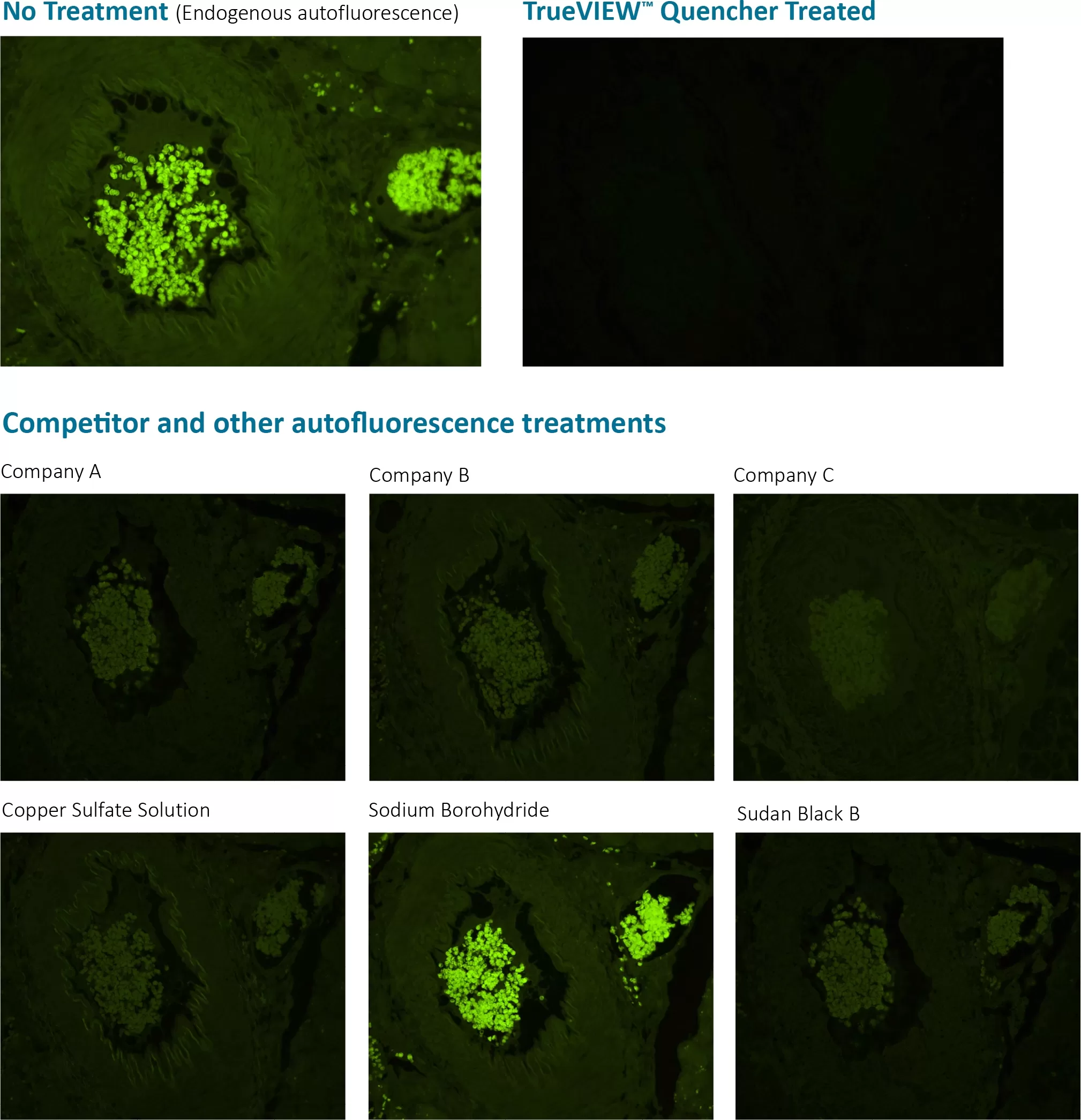
Comparison Between Vector TrueVIEW Reagent and Other Autofluorescence Reducing AgentsWe compared the effectiveness of Vector TrueVIEW quenching action in parallel with other commercially available autofluorescence reducing products and “home brew” reagents, on serial sections of formalin-fixed, paraffin-embedded human pancreas visualized using a standard fluorescein (green) filter. No specific immunofluorescence staining was conducted. The images below highlight our results. All images were acquired under identical conditions (including microscope objective and exposure times).
|
 Powered by Bioz © 2023
Product FAQs
Why is the TrueVIEW™ Autofluorescence Quenching reagent applied after completion of the IF assay and not at the start of the procedure?
During product development, applying the TrueVIEW™ Quenching reagent at the end of our standard IF procedure yielded the most optimal reduction in autofluorescence signal. The reagent is retained on the tissue at the time of mounting, allowing for extended quenching action, with little to no effect on the specific fluorescent signal. However, in applications with very brief staining procedures, such as a primary antibody directly conjugated with a fluorophore, application of TrueVIEW™ Quenching reagent may be just as effective at the start of the procedure.
Do you have any published references describing the use of TrueVIEW Autofluorescence Quenching Kit?
Yes, there are a number of published references describing the use of TrueVIEW Autofluorescence Quenching Kit in the scientific literature. Please refer to a partial list of these publications in the Technical Information section of the product detail page for SP-8400.
Is TrueVIEW effective against lipofuscin derived autofluorescence?
The working solution of TrueVIEW works in an electrostatic manner to greatly reduce or eliminate fluorescence in tissue sections induced through the use of an aldehyde based fixative. TrueVIEW is also effective at reducing fluorescence from tissue components such as collagen, elastin and red blood cells. TureVIEW does not work to reduce autofluorescence due to lipofuscin.
How long can I store the TrueVIEW working solution?
Once made up, the working solution of TrueVIEW can be stored either on the bench top or in the fridge (2-8C) fro about 48 hours (2 days) withut loss of activity or function. Following this time we would suggest discarding unused working solution and make fresh solution as required.
My tissue sections turned blue when I applied TrueVIEW. Is this supposed to happen?
Yes, the working solution is a blue color that does “stain” the tissue section blue. This indicates an active and appropriate chemical reaction is occurring. The blue stain on the section does not fluoresce and does not interfere with the immunofluorescence application.
What mounting media can I use with TrueVIEW?
Both TrueVIEW products, SP-8400 and SP-8500, are supplied with 2 mL of VECTASHIELD Vibrance antifade mounting media. We have found that the mounting media does play a crucial role in maintaining the high signal to noise ratio when using TrueVIEW. VECTASHIELD Vibrance is included therefore as a critical component of the kit and it is recommended to use the media supplied for optimal results. At this time we do not have sufficient data to confidently recommend the use of other vendors mounting media with TrueVIEW. Substitution of VECTASHIELD Vibrance with another mounting media may result in less than satisfactory results.
For the wash step after applying the TrueVIEW quenching reagent, can I use buffers other than PBS or detergents?
No, we have found that the TrueVIEW reagent lifts off the tissue using TBS or HEPES buffer. Detergents are incompatible.