
Selvakumar Subbian
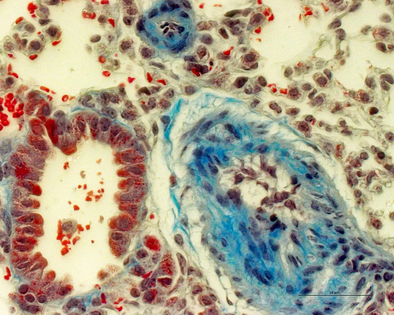
Mouse lung infected with Mycobacterium tuberculosis stained with Masson’s Trichrome to visualize collagen deposition, fibrosis and tissue remodeling. The main structures shown are a bronchus and a pulmonary vessel. Nuclei are stained brown, cytoplasm and erythrocytes are stained red and collagen is stained blue. Image captured by Dr. Selvakumar Subbian, New Jersey Medical School, Rutgers University. December 2020
Products used: H-6100, SP-1800-7, H-5000-60