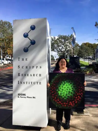
Catherine Cheng, The Scripps Research Institute, USA.

Catherine Cheng
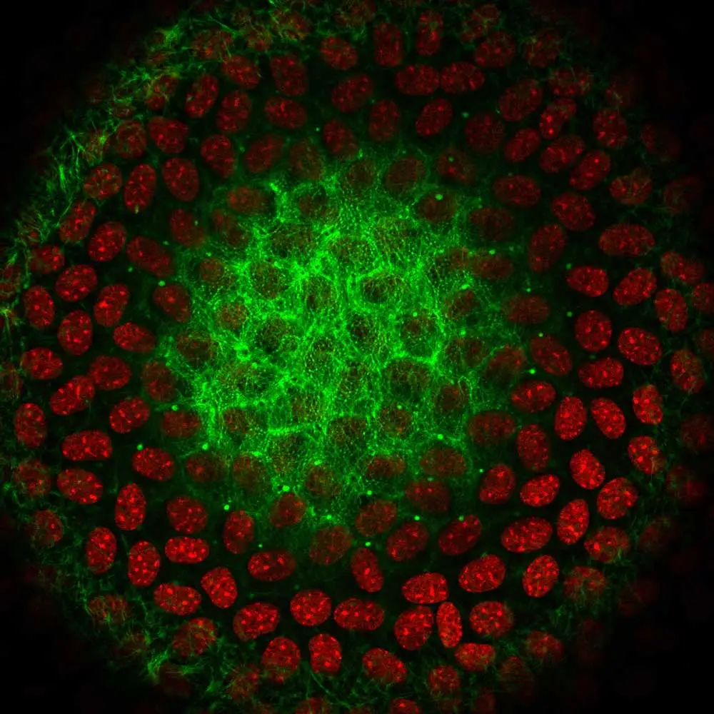
The lens is a transparent and avascular organ in the front of the eye that is responsible for focusing light onto the retina in order to transmit a clear image. A monolayer of epithelial cells covers the anterior hemisphere of the lens, and the bulk of the lens is made up of fiber cells. Epithelial cells near the anterior pole are cuboidal in shape and contain diverse actin filaments.1 This image shows one optical section of a whole mouse lens stained with phalloidin (F-actin, green) and DAPI (nuclei, red). The basal surfaces of these anterior epithelial cells contain actin stress fibers and actin filament-rich lamellipodia (cells in the outermost ring). On the lateral surfaces, anterior epithelial cells are polygonal in shape with actin cortical fibers and sequestered actin bundles (cells in second ring from the outside with bright green F-actin puncta). Apical domains of the anterior epithelial cells contain polygonal arrays of actin filaments11 (cells in the middle). These geodesic domes of actin are hypothesized to flatten in order to maintain epithelial cell integrity during lens shape changes (accommodation) so you can see near objects clearly.
References:
1. Cheng C, Nowak RB, Fowler VM. The lens actin filament cytoskeleton: Diverse structures for complex functions. Experimental eye research 2016.
2. Liou W, Rafferty NS. Actin filament patterns in mouse lens epithelium: a study of the effects of aging, injury, and genetics. Cell motility and the cytoskeleton 1988; 9:17-29.
3. Weber GF, Menko AS. Actin filament organization regulates the induction of lens cell differentiation and survival. Developmental biology 2006; 295:714-29.
4. Bassnett S. Three-dimensional reconstruction of cells in the living lens: the relationship between cell length and volume. Experimental eye research 2005; 81:716-23.
5. Rafferty NS. Lens Morphology. New York: Dekker, 1985.
6. Rafferty NS, Scholz DL. Actin in polygonal arrays of microfilaments and sequestered actin bundles (SABs) in lens epithelial cells of rabbits and mice. Current eye research 1985; 4:713-8.
7. Rafferty NS, Scholz DL. Polygonal arrays of microfilaments in epithelial cells of the intact lens. Current eye research 1984; 3:1141-9.
8. Rafferty NS, Scholz DL. Comparative study of actin filament patterns in lens epithelial cells. Are these determined by the mechanisms of lens accommodation? Current eye research 1989; 8:569-79.
9. Rafferty NS, Scholz DL, Goldberg M, Lewyckyj M. Immunocytochemical evidence for an actin-myosin system in lens epithelial cells. Experimental eye research 1990; 51:591-600.
10. Scholz DL, Rafferty NS. Immunogold-EM localization of actin and vimentin filaments in relation to polygonal arrays in lens epithelium in situ. Current eye research 1988; 7:705-19.
11. Yeh S, Scholz DL, Liou W, Rafferty NS. Polygonal arrays of actin filaments in human lens epithelial cells. An aging study. Investigative ophthalmology & visual science 1986; 27:1535-40.
Products used: H-1200-10

This image was captured by Dr. Catherine Cheng The Scripps Research Institute, USA.
8. Rafferty NS, Scholz DL. Comparative study of actin filament patterns in lens epithelial cells. Are these determined by the mechanisms of lens accommodation? Current eye research 1989; 8:569-79.
9. Rafferty NS, Scholz DL, Goldberg M, Lewyckyj M. Immunocytochemical evidence for an actin-myosin system in lens epithelial cells. Experimental eye research 1990; 51:591-600.
10. Scholz DL, Rafferty NS. Immunogold-EM localization of actin and vimentin filaments in relation to polygonal arrays in lens epithelium in situ. Current eye research 1988; 7:705-19.
11. Yeh S, Scholz DL, Liou W, Rafferty NS. Polygonal arrays of actin filaments in human lens epithelial cells. An aging study. Investigative ophthalmology & visual science 1986; 27:1535-40.
Products used: H-1200-10

