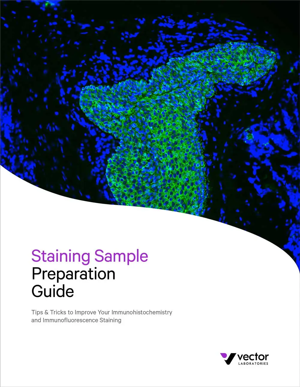
Staining Sample Preparation Guide
Tips & Tricks to Improve Your Immunohistochemistry and Immunofluorescence Results
Sitting at the microscope at the end of an IHC or IF experience can spark feelings of amazement as you uncover the result you’ve been patiently waiting for. Getting to this point requires careful planning and execution to ensure the best results with appropriate tissue morphology and low background. Tissue sample preparation includes multiple steps, and many of them offer different choices – paraffin embedding versus cryopreservation, section thickness, and antigen retrieval buffer, among others. In this guide, we will walk you through key decisions and troubleshooting issues to empower you to ask the right questions and optimize your protocols
What’s inside:
- Considerations for sample preservation and storage
- Tips to get the most out of your sectioning
- Advice for deparaffinization and rehydration
- Suggestions for antigen retrieval
- How to save time and tissue with hydrophobic barrier pens
Please submit the following form to request a copy of this guide. Note that fields marked
with an asterisk * are required.


