Reactivity Amine | |
Unit Size 1 Kit | |
Storage Instructions 2° – 8°C – Do Not Freeze | |
Applications In situ hybridization, In Situ Proximity Ligation | |
Conjugate Antibody | |
Label Digoxigenin |

ChromaLINK® Digoxigenin One-Shot™ Antibody Labeling Kit
The ChromaLINK Digoxigenin One-Shot Antibody Labeling Kit provides convenient, consistent, and measurable digoxigenin labeling of 100 µg of antibody. Each kit contains ChromaLINK Digoxigenin, which incorporates a novel UV-traceable chromophore in the linker arm to enable reproducibility in your antibody labeling process. With a simple, non-destructive UV scan you can now quantify digoxigenin incorporation to ensure reproducible incorporation of the optimal number of haptens per antibody. Each ChromaLINK One-Shot kit contains everything needed to to label your antibody: buffers, reagents, desalting columns, and an easy-to-follow protocol and online DIG incorporation calculator.
$340.00
| SKU | Unit Size | Price |
|---|---|---|
Select a unit size:
How do I request a quote or bulk pricing?
Kit Components
|
Technical Information Product DescriptionThe ChromaLINK Digoxigenin One-Shot antibody labeling kit is specifically designed to incorporate a digoxigenin label into a single 100 microgram quantity of antibody resuspended in 100 μl of buffer. This kit comes complete with all the necessary components to label and rapidly quantify incorporated digoxigenin from a small quantity of antibody in about 90 minutes. Digoxigenin has long been used as an alternative hapten reporter system to the streptavidin/biotin system (1). This system is based on a steroid isolated from the blossoms and leaves of the plant Digitalis purpurea, the only known natural source of digoxigenin. High affinity antibodies raised against digoxigenin thus exhibit virtually no cross-reaction with other biological molecules found in higher organisms. Both the specificity and sensitivity of this “parallel” hapten-reporter system often gives digoxigenin a major advantage over biotin-based detection systems. In more recent times, this reporter system has also been used in conjunction with the biotin-streptavidin system to detect multiplexed antigens (2). Unlike the biotin/streptavidin system, the digoxigenin label has never had a method for directly quantifying incorporation, until now. The labeling process used in the ChromaLINK Digoxigenin One-Shot kit is based on a patented UV-traceable linker called Sulfo-ChromaLINK Digoxigenin (Figure 1). This advanced digoxigenin labeling reagent contains an aromatic water-soluble N-hydroxy-sulfosuccinimidyl ester functional group (a), which efficiently modifies protein lysine residues in a phosphate buffer system. The linker also possesses an embedded bis-aryl hydrazone structure (b), which forms the compound’s UV-traceable chromophore. This traceable signature enables the non-destructive and direct quantification of digoxigenin (c), attached to the antibody. These diverse chemical elements are linked together through a long-chain PEG3 spacer (d), which preserves antibody-digoxigenin binding affinity while simultaneously maintaining antibody solubility. Benefits and FeaturesThe ChromaLINK Digoxigenin One-Shot kit is a convenient, cost-effective way of incorporating digoxigenin into a small quantity (100 μg) of an antibody. After incorporation, digoxigenin is rapidly quantified using a non-destructive UV-scan (220-400 nm). The kit features high antibody recoveries (50-80%) with consistent digoxigenin incorporation. The degree of incorporation or molar substitution ratio (MSR) ranges from 2-8 digoxigenin molecules per antibody when used as directed. The ChromaLINK Digoxigenin One-Shot kit can be used to label a diverse group of antibodies including all mammalian IgG, IgE, and avian IgY molecules. The ChromaLINK Digoxigenin One-Shot kit offers a robust method of both labeling and quantifying incorporated digoxigenin with a simple protocol. This “double-feature” provides researchers with greater confidence, consistency and reproducibility in all downstream applications. Procedure Diagram
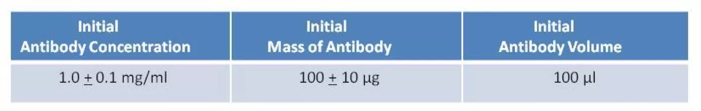
Figure 2. Illustration of the ChromaLINK Digoxigenin One-Shot antibody labeling procedure. Process SummarySample Preparation: adjust antibody to 1 mg/ml in 100 μl buffer Sample Analysis: confirm antibody concentration using a spectrophotometer First Buffer Exchange: equilibrate spin columns & buffer exchange antibody Sample Analysis: reconfirm recovered antibody concentration on a spectrophotometer Digoxigenin Labeling: label antibody with ChromaLINK Digoxigenin linker Second Buffer Exchange: remove excess labeling reagent with spin column Digoxigenin Incorporation: quantify incorporation using a spectrophotometer Important Labeling ParametersThe ChromaLINK Digoxigenin One-Shot antibody labeling kit is specifically designed to incorporate digoxigenin into a single 100 microgram quantity of antibody resuspended in 100 μl of a suitable buffer as indicated in Table 1.
Table 1. Initial starting conditions required for the ChromaLINK Digoxigenin One-Shot procedure. The kit provides consistent and reliable incorporation of digoxigenin by controlling the following reaction variables:
Critical to digoxigenin incorporation is the ability to accurately determine an antibody’s initial concentration in a non-destructive manner with full recovery of the sample. The ChromaLINK Digoxigenin One-Shot procedure employs a spectral scan (220-400 nm) rather than a single wavelength measurement @ 280 nm to accomplish this task. Commercial antibodies often contain preservatives or other additives capable of masking or distorting the intrinsic UV spectrum of a sample. As a consequence, additives make it more difficult to accurately estimate protein concentration using a single wavelength (i.e. A280) measurement. A scan however, provides greater information and assurance that the antibody’s concentration is correct since a spectrum often reveals the presence of interfering additives. Commonly used additives may include preservatives such as sodium azide, thimerosal, protein stabilizers such as *BSA or *gelatin, or small molecule additives such as glycine. *Note-protein carriers such as BSA or gelatin must be removed before labeling can proceed. |
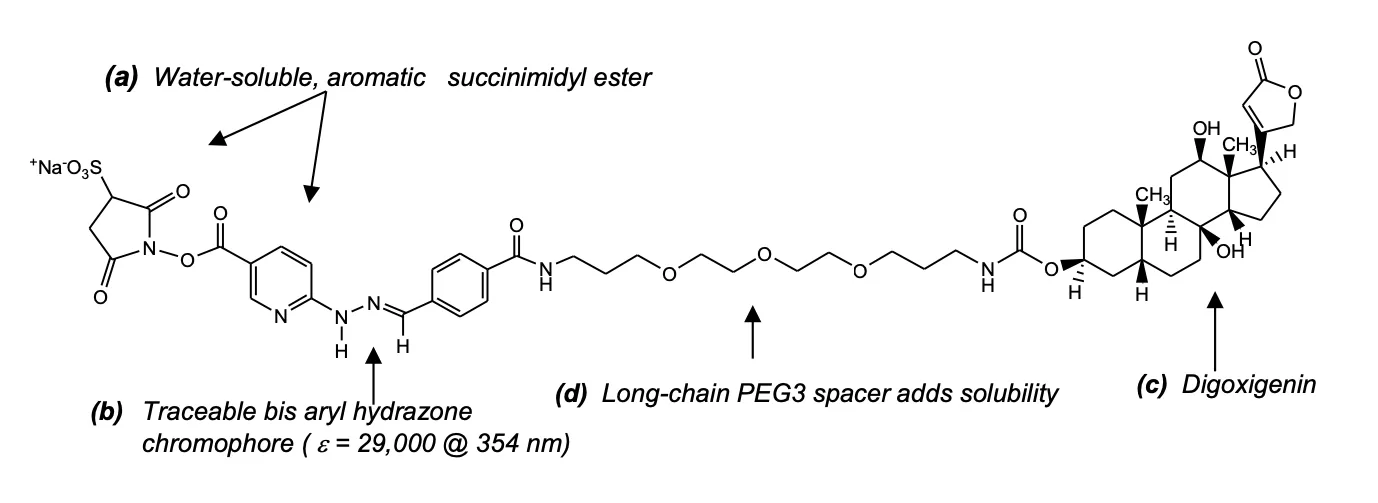 Figure 1: Structure of ChromaLINK Digoxigenin C52H67N6NaO17S; M.W. 1103.17
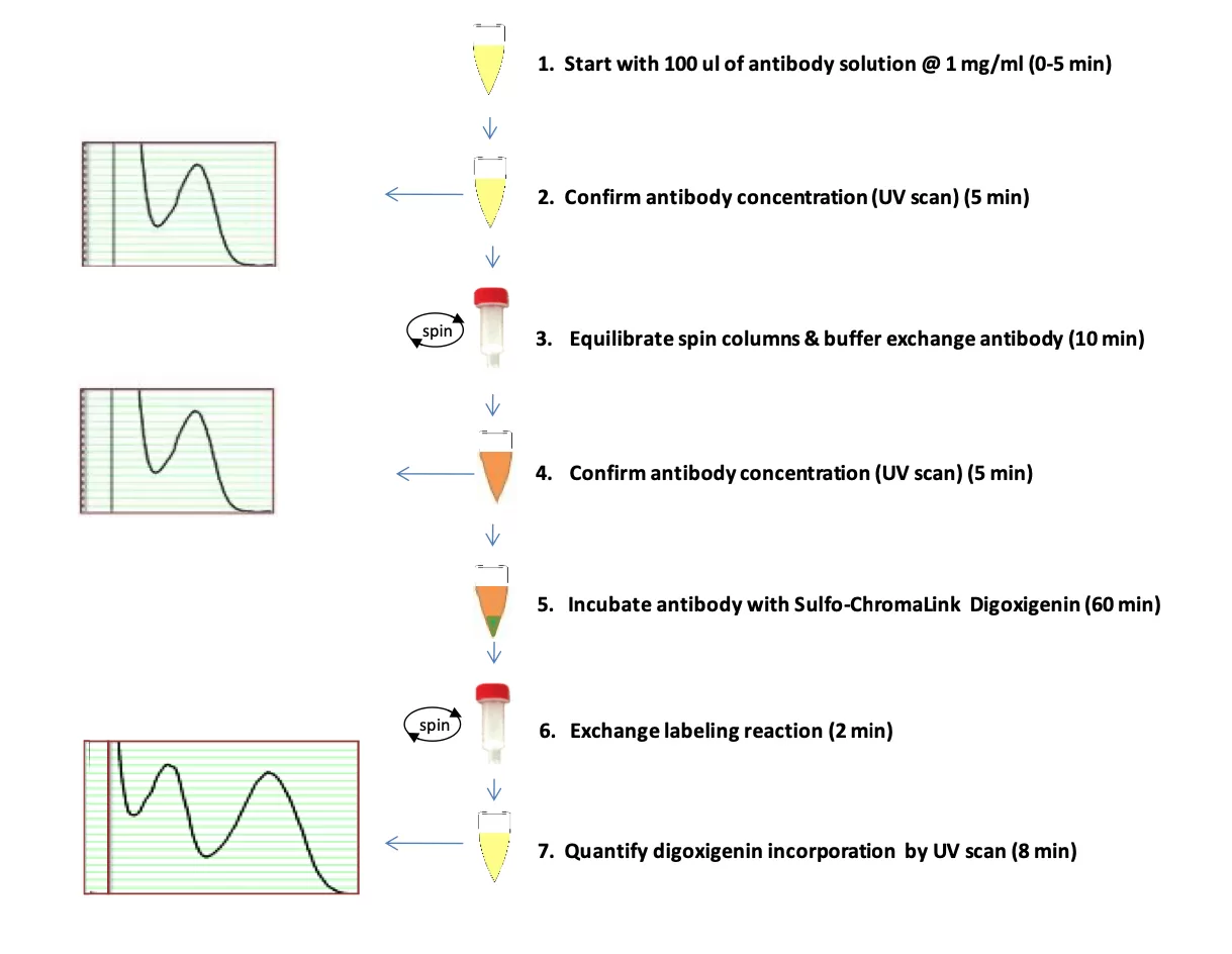
Legal Notice SoluLINK® Bioconjugation For Research Use Only. Not for use in diagnostic procedures. For additional licensing restrictions, please see the license agreement at vectorlabs.com/solulink-research-license. Products are for research use only, not for use in diagnostic or therapeutic procedures or for use in humans. Products are not for resale without express written permission of Seller. No license under any patent or other intellectual property right of Seller or its licensors is granted or implied by the purchase unless otherwise provided in writing. |