
Vector Laboratories is closed for the President’s Day on Monday, February 19th. We will be back in the office on Tuesday, February 20th.
We will respond to emails upon our return. Have a wonderful day.
Menu
Vector Laboratories is closed for the President’s Day on Monday, February 19th. We will be back in the office on Tuesday, February 20th.
We will respond to emails upon our return. Have a wonderful day.
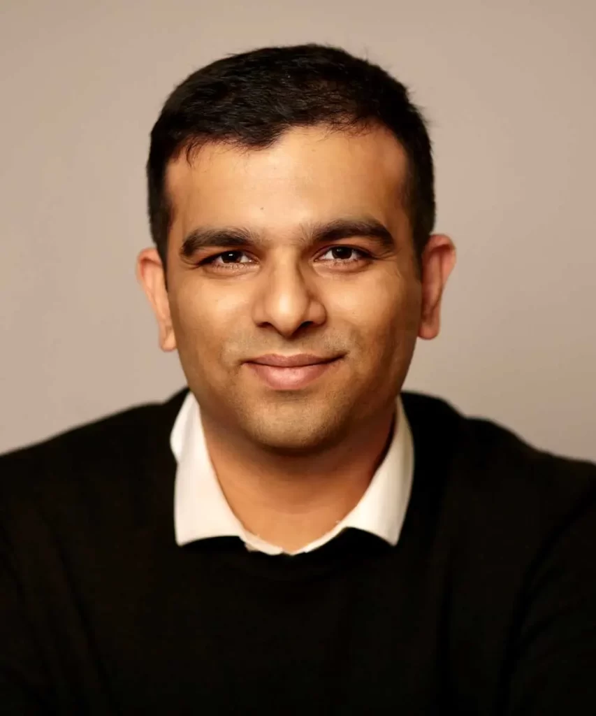
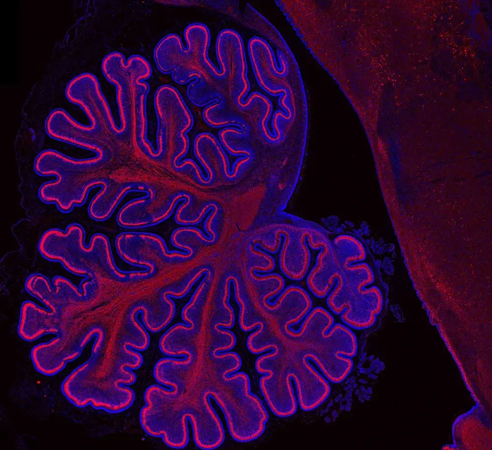
Parthiv Haldipur is a researcher focused on developmental neuroscience with a quest to better understand brain development and its effects from conception throughout adulthood. Parthiv traces his earliest interest in science to a wonderful science teacher in fourth grade who introduced his class to the concept of how a surface area can be increased through folding using the human biology of the intestine as an example. Parthiv was fascinated that the extra-long intestinal tract was able to fold upon itself to fit into the abdominal cavity and that the mass of fingerlike projections of the intestinal wall dramatically expanded the surface area. When his parents gave him a microscope for his tenth birthday, no one could portend how this basic tool would pique his curiosity and serve as a beacon over the course of his career. And while the microscopes he uses now have more capabilities than that childhood toy, he believes that the best discoveries are made under the microscope.
“I am very often astounded by how logical biological patterns are. When you look at the process of how cells are arranged in the brain, it may seem random. But when you identify those patterns, it is incredible how everything makes sense. Progenitors and neurons are arranged in a specific manner so that ultimately the brain can function efficiently. And, if you disturb that arrangement, things can go awry…some people just think of the brain as a tangled mass of neurons and that the organization doesn’t matter…But there’s a reason why brain cells are arranged in a particular manner.”
Parthiv credits all the fantastic biology educators throughout his life as positive influencers, from elementary and high school teachers to university professors and research mentors. As an undergraduate at St. Joseph’s College (Bangalore University, Bengaluru, India), he studied microbiology, chemistry, and zoology. He discovered his passion for neuroscience and developmental biology during his master’s education at Maharaja Sayajirao University of Baroda (Vadodara, India). He later earned a PhD at the National Brain Research Centre (Gurgaon, India) in developmental neuroscience. In 2012, he began a postdoctoral fellowship studying cerebellar development and disease at Seattle Children’s Research Institute (Washington, US). He was promoted to Research Scientist in 2019 and still works in that same lab at Seattle Children’s, continuing his research and looking forward every day to his time at the bench.
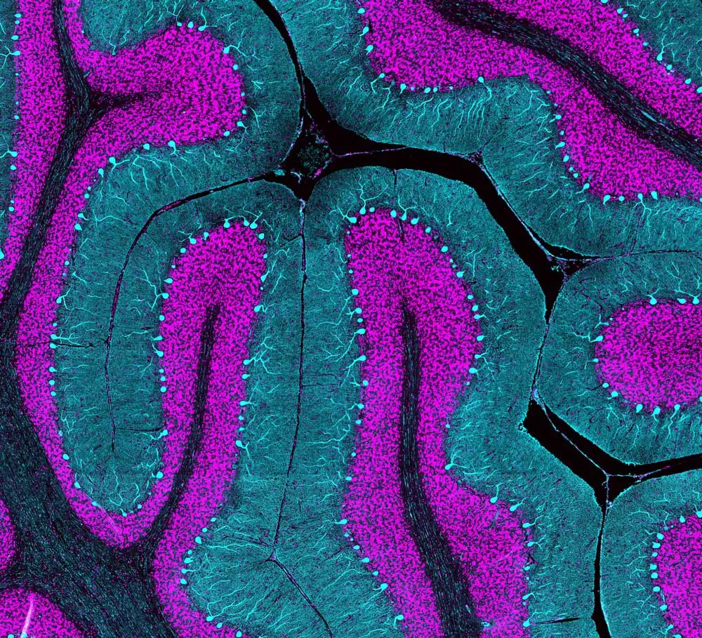
“While some of the differences observed between human and animal models are subtle, the rest are striking. When we began looking at the developing human cerebellum, we found several significant differences in histological and pathological patterns between the mouse and human, and in fact between non-human primates and humans, as well. These differences have become significant in the context of human disease.”
Working under the expectation that brain development was fully conserved among species, neuroscientists have relied upon mouse and non-primate species as viable animal models of human disease. However, recent studies in closely related human and non-human primates have unveiled significant developmental differences, evidenced by histological and pathological pattern data. In 2015, Parthiv and his colleagues were able to gain invaluable access to human cerebellum samples for research purposes, ethically acquired from autopsies in collaboration with pathologists around the world. Since each sample can only represent a single snapshot in time, it remains difficult to study a human development continuum. Instead, developmental clues are revealed one at a time until enough data is available to solve each puzzle.
“I like doing my own microscopy because it’s under the microscope where you make discoveries. In 2019, we published a paper in Science where we described histopathological differences between the developing human and mouse cerebella. Many of those differences were identified under the microscope. For me, the microscope is like what a stethoscope is to a doctor. I spend most of my time in the microscope room, and that is an essential part of my life as a researcher.”
Because developmental issues in the brain early in life can be linked to brain defects and diseases that arise later, Parthiv knows that it is essential to learn as much as possible. He studies abnormal development of the cerebellum involved in the rare, congenital defect or malformation known as Dandy-Walker Malformation. He also is examining how developmental origins might be implicated in medulloblastomas, highly malignant pediatric brain tumors. He hopes to see the knowledge gathered today in laboratories culminate into better translational medicine diagnoses, treatments, and outcomes in the future.
“Multitasking has been a bit challenging. As you gain more experience and get promoted, your responsibilities increase. Then you have to balance benchwork with funding acquisition… I like being a Research Scientist because I want to do benchwork for the rest of my life as a researcher… I want to continue making those discoveries.”
Having benefitted from great teachers and mentors who provided different forms of mentoring throughout his life, Parthiv finds satisfaction in passing along the skills and knowledge he acquired along the way to the next generation. He sees that students in the process of learning end up asking insightful questions that might not have otherwise crossed his mind. This kind of shift, which temporarily reverses the flow of wisdom from the student to the mentor, can set the stage for the development of an enduring and mutually beneficial relationship. For the students who see the joy he finds under the microscope, it very well could lead them into their own lifelong journey, filled with wonder and discovery.


Stay in the Loop. Join Our Online Community
Together we breakthroughTM

©Vector Laboratories, Inc. 2024 All Rights Reserved.