
Vector Laboratories is closed for the President’s Day on Monday, February 19th. We will be back in the office on Tuesday, February 20th.
We will respond to emails upon our return. Have a wonderful day.
Menu
Vector Laboratories is closed for the President’s Day on Monday, February 19th. We will be back in the office on Tuesday, February 20th.
We will respond to emails upon our return. Have a wonderful day.
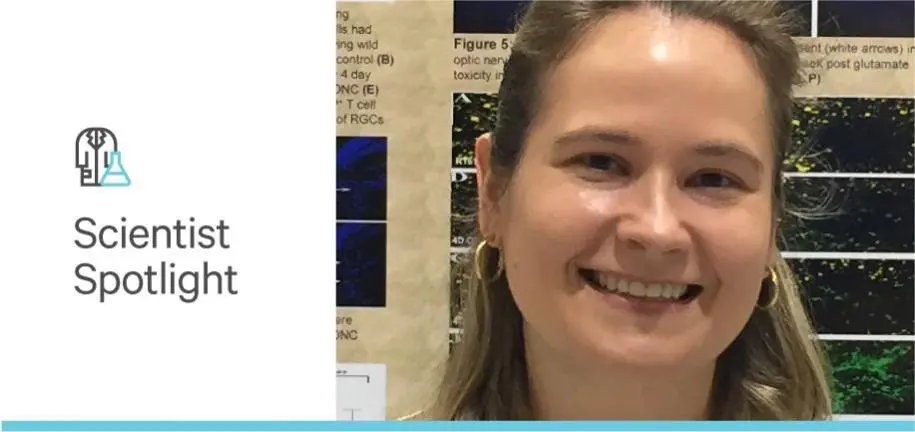

Jennifer Kielczewski (MS, PhD) has been at the National Institutes of Health (NIH) since 2011. After completing her postdoctoral work there, focusing on the retina in the Lab of Immunology, she began her current staff scientist position in 2016 at the Biological Imaging Core (BIC) within the NIH National Eye Institute (NEI). The mission of the NEI is to promote healthy vision by supporting and conducting basic research on the eye. The BIC primarily images species from mouse, but some analyses include studies that use human retinas from eye bank donations. The BIC provides services that include imaging the retina, staining for specific proteins and protein localization, quantifying information from confocal images, assessing genetic defects in animal models and diseased models for glaucoma, diabetic retinopathy, age-related macular degeneration (AMD), and other ocular disorders. Especially promising is the translational potential of stem cell research to restore vision as these types of therapies move closer to clinical trials.
“When postdocs come into the Institute, we have to train them from all levels of experience. If they have some microscopy experience, they’re going to learn very quickly and for others it can take more time to master imaging, but we try to make sure everyone collects images that of high quality”.
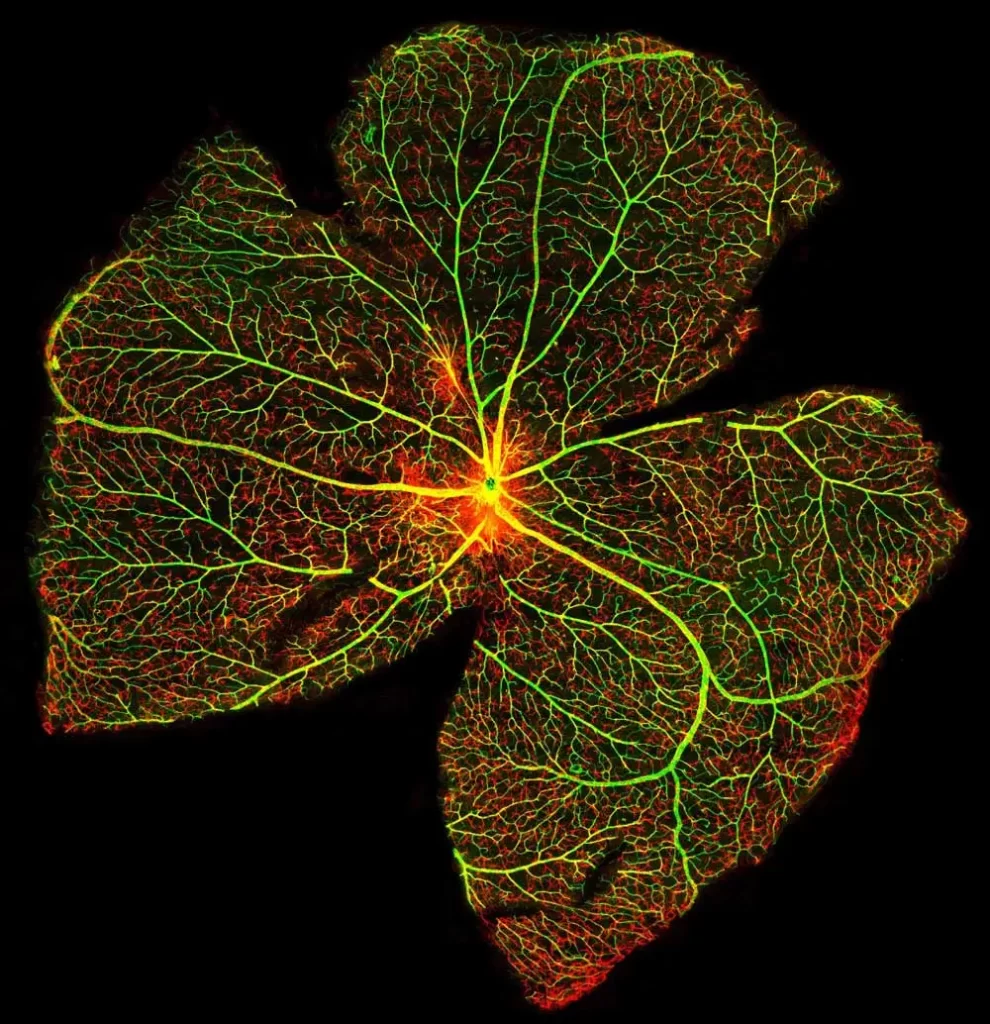
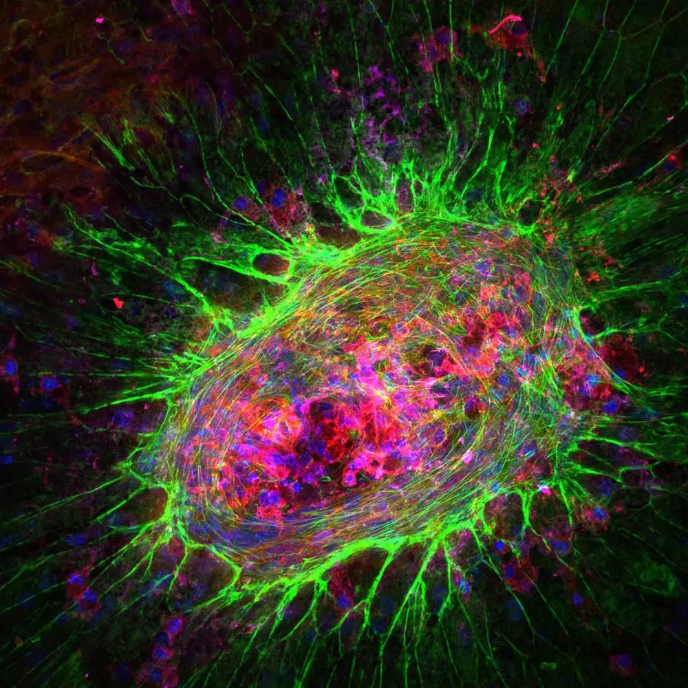
Jennifer’s service role in the BIC involves not only providing hands-on training to approximately 50 trainees as they rotate in and out of NEI but also maintaining the microscopy equipment, offering imaging and analysis advice as needed, and participating in collaborative projects within the NIH and with outside institutions. Much of the retinal imaging that relies on her consultation involves immune cells or other areas of immunology and immunopathology. The BIC selects antibodies and other imaging reagents known to help ensure robust signals, including ones that Jennifer used in her graduate and post-graduate research. These include reliable photoreceptor stains like carbohydrate-specific peanut agglutinin (PNA), our hydrophobic barrier ImmEdge Pen to ensure that the solution stays on the slides during the staining process, and VECTASHIELD mounting media to maintain fluorescence intensity and prevent signal fading.
“My background as a postdoc involved a lot of immunology work in the retina. As a result, I do look at a lot of retinas that are diseased with inflammation, that have immune cells in them or have some other type of immunological pathology.”
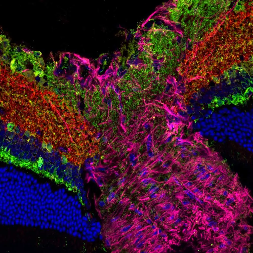
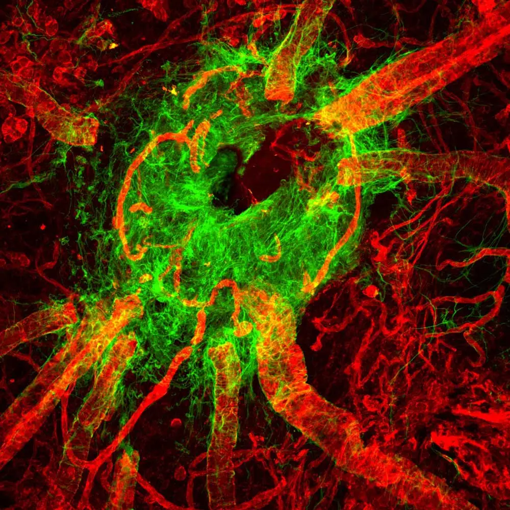
The primary objective of the BIC is to provide imaging services, experimental design and workflow guidance and support to researchers working on eye specimens. High demand for BIC services is evidenced by the fact that their microscopes are nearly booked every day. The facility offers a broad range of imaging options for in vivo and in vitro applications including epifluorescent, confocal and super-resolution microscopy. Jennifer and the dedicated BIC team utilize their expertise to leverage resources available at the facility to ensure each projects objectives and investigator’s research needs are met. Generating the images and seeing the “really gorgeous” end results, are some of Jennifer’s favorite aspects of working at BIC.


Stay in the Loop. Join Our Online Community
Together we breakthroughTM

©Vector Laboratories, Inc. 2024 All Rights Reserved.