
Vector Laboratories is closed for the President’s Day on Monday, February 19th. We will be back in the office on Tuesday, February 20th.
We will respond to emails upon our return. Have a wonderful day.
Menu
Vector Laboratories is closed for the President’s Day on Monday, February 19th. We will be back in the office on Tuesday, February 20th.
We will respond to emails upon our return. Have a wonderful day.


Dr. Enrico Radaelli, DVM, PhD, DECVP has been the Technical Director of the Comparative Pathology Core (CPC) since the spring of 2017. Founded in 2017 by Enrico’s colleague Amy Durham, the Faculty Director of the CPC is part of the Abramson Cancer Center at the University of Pennsylvania Veterinary School in Philadelphia. Enrico supervises the day-to-day scientific activities for a small core team of fellows and technicians within the Department of Pathobiology at the Ryan Veterinary Hospital. While the CPC team works on a few collateral research projects, 90 percent of the services that they provide are in support of the research endeavors of over a hundred customers, including those on the Penn campus, other academic researchers, and private organizations in the United States and Europe. The core laboratory environment is very dynamic, enabling a new discovery almost daily, but as a result it can be challenging to keep up with. New protocols and techniques need to be validated before the CPC can offer those services, and this means that the core needs access to a variety of animal and human samples. Finding the proper samples is not always easy but established funding systems can help.
“… unlike in a hospital, we are just working with human tissues where you have your kit and it’s working for all the samples. We are in a situation where today we have fish, tomorrow we have mouse, and the other day we had monkey …”
Enrico, a certified veterinary pathologist trained in Italy and Belgium, is motivated by the potential of translational research in that it sits at the cusp between basic research and clinical research and his unique comparative pathology perspective helps improve interpretation of the findings from the core’s services. In addition to the services the team provides, Enrico has undertaken the role of educating clientele – not only about the CPC’s experimental pathology services that include processing tissue, immunohistochemical staining, in situ hybridization, digital image analysis, and more – but also how to create a robust, humane, optimized animal model design. This kind of consultation can include how to use animal models to test drugs and other experimental conditions, which animals are relevant in the study of specific human diseases, how to minimize unnecessary procedures and animal suffering, and how comparative pathology affects translating the animal model results into understanding the effects in humans.
“The general concept is that, you are a vet–you know about the companion animals and lab animals. But actually, our discipline, especially in pathology, is really focused on the comparative aspects of being able to go from the animal to human, and over this translational process, trying to create knowledge that is relevant for the clinical setting.”
While the Abramson Cancer Center and University of Pennsylvania Veterinary School provide advertising support for the very busy seven-member core lab services team, Enrico senses that the best advertisers are the satisfied customers who share their word-of mouth recommendations. He is proud of the team and the role he plays in it, the level of service that they provide, how they meet the deadlines of the investigators with reasonable turnaround times, and that they have provided a lot of data for many publications. Yet his main motivation is rooted in his curiosity in comparative veterinary pathology, which enables leveraging animal models to better understand what’s happening in humans and translating that knowledge into the clinic.
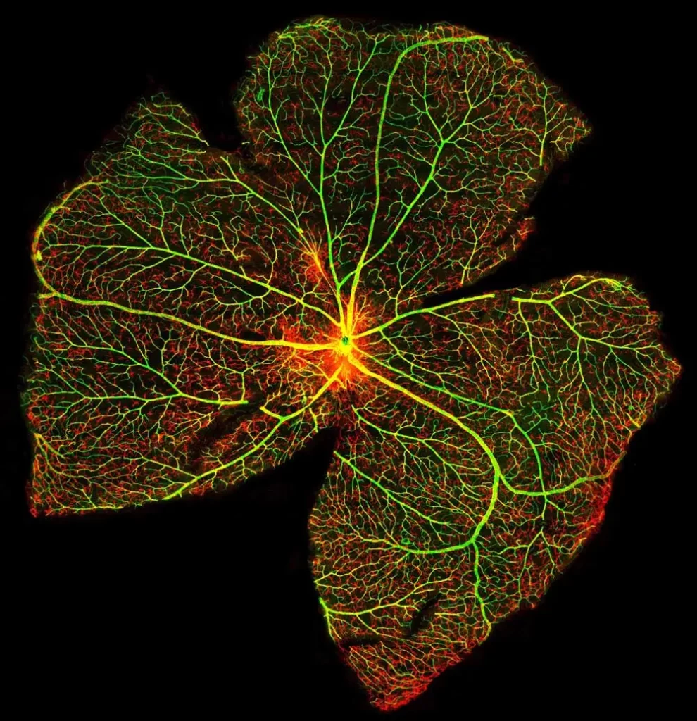
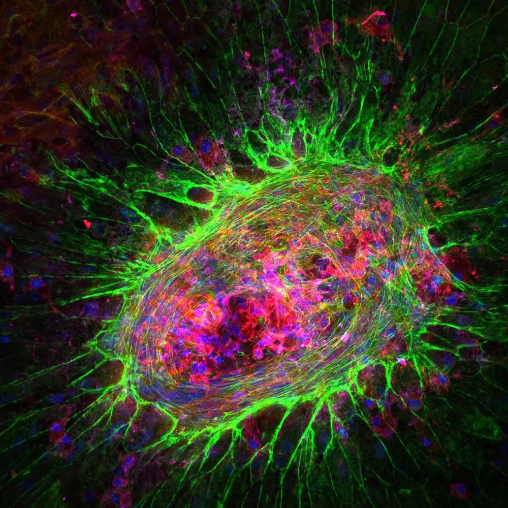
Jennifer’s service role in the BIC involves not only providing hands-on training to approximately 50 trainees as they rotate in and out of NEI but also maintaining the microscopy equipment, offering imaging and analysis advice as needed, and participating in collaborative projects within the NIH and with outside institutions. Much of the retinal imaging that relies on her consultation involves immune cells or other areas of immunology and immunopathology. The BIC selects antibodies and other imaging reagents known to help ensure robust signals, including ones that Jennifer used in her graduate and post-graduate research. These include reliable photoreceptor stains like carbohydrate-specific peanut agglutinin (PNA), our hydrophobic barrier ImmEdge Pen to ensure that the solution stays on the slides during the staining process, and VECTASHIELD mounting media to maintain fluorescence intensity and prevent signal fading.
“My background as a postdoc involved a lot of immunology work in the retina. As a result, I do look at a lot of retinas that are diseased with inflammation, that have immune cells in them or have some other type of immunological pathology.”
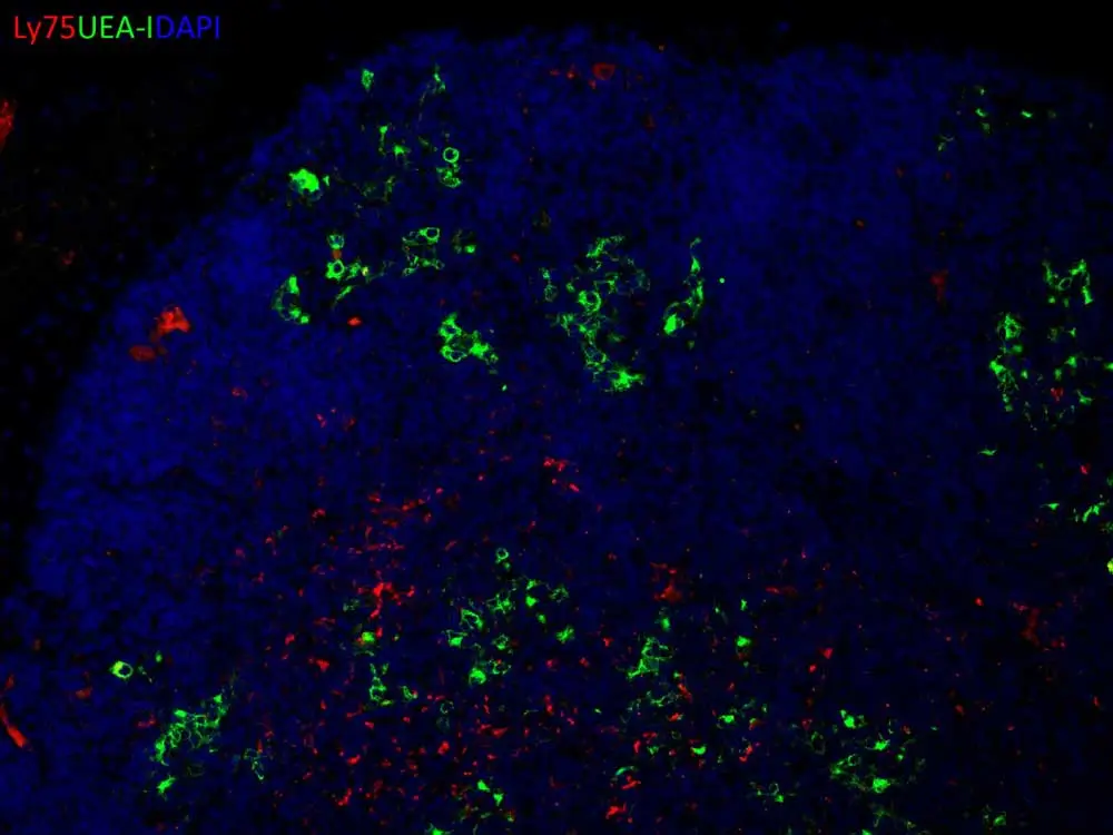
Duplex immunofluorescence staining of a thymic lymphoma in a genetically engineered mouse. The combination of Ly75 antibody staining (red) and UEA-I lectin labeling (green) allows the study of the cortical and medullary thymic epithelium, respectively, during lymphomagenesis. The following lectin product from Vector has been used to label the medullary epithelial compartment: Fluorescein labeled Ulex Europaeus Agglutinin I (UEA-I), Catalog Number: FL-1061.
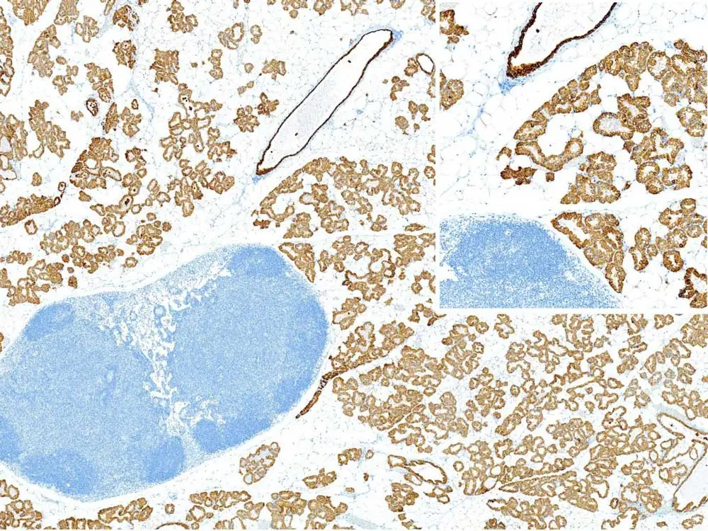
Keratin 8 (Rat monoclonal antibody DSHB, Catalog # TROMA-I) immunoperoxidase staining of the mammary gland in a female mouse at the end of pregnancy. Diffuse and intense immunoreactivity is evident in the luminal epithelium of the acinar and ductal structures. The following kit from Vector has been used to develop this staining: ImmPRESS® HRP Goat Anti-Rat IgG, Mouse adsorbed (Peroxidase) Polymer Detection Kit, Catalog Number: MP-7444.


Stay in the Loop. Join Our Online Community
Together we breakthroughTM

©Vector Laboratories, Inc. 2024 All Rights Reserved.