
Vector Laboratories is closed for the President’s Day on Monday, February 19th. We will be back in the office on Tuesday, February 20th.
We will respond to emails upon our return. Have a wonderful day.
Menu
Vector Laboratories is closed for the President’s Day on Monday, February 19th. We will be back in the office on Tuesday, February 20th.
We will respond to emails upon our return. Have a wonderful day.
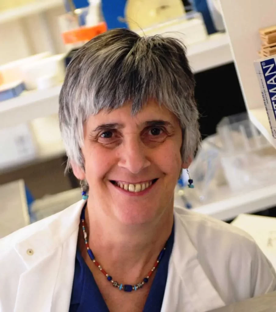
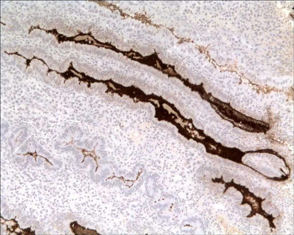
Carolyn Jones embarked on her academic science career as a mature student in 1967, continues to work in the field of electron microscopy and lectin histochemistry, and describes herself as a microscopist and glycobiologist. She now works mainly on the glycosylation of reproductive tissues, particularly animal placentae which have ranged from that of mouse to the elephant. Though officially retired in 2008, she notes that nothing has really changed while she continues her research in an honorary capacity. If you look at her CV, it may look typical, but her journey has been anything but typical. With an unassuming personality, she may seem to gloss over her accomplishments but, indeed, she founded the science of reproductive glycogenetics, revealing the existence of a feto-maternal glycocode unique to each species, and involving the specific interaction of sugars on placental surfaces with those on the apposing maternal endometrium.
Carolyn also has an artistic side that affected her academic training from the start, as she originally wanted to be a medical illustrator, and that has translated well when, instead, she went into science. She is totally hooked on the beauty unveiled by light and electron microscopy. She describes her career pathway as one filled with serendipity–from the happenstance timing of open positions that matched her interest—to chance collaborations initiated on a train ride—to the opportunities she took because her job responsibilities were ill-defined—all these became intricately woven together into an extraordinary life.
“After I lost income from providing services in electron microscopy, the head of my department began supporting me financially, paying for consumables that I needed to carry on my research. Basically, from the day I retired in 2008 and my position as Research Fellow became honorary, nothing changed. I still went in to work and published. It’s just that there’s no pressure to have a 9-to-5 working day, so I go in when I like and come home when I like, and it’s lovely.”
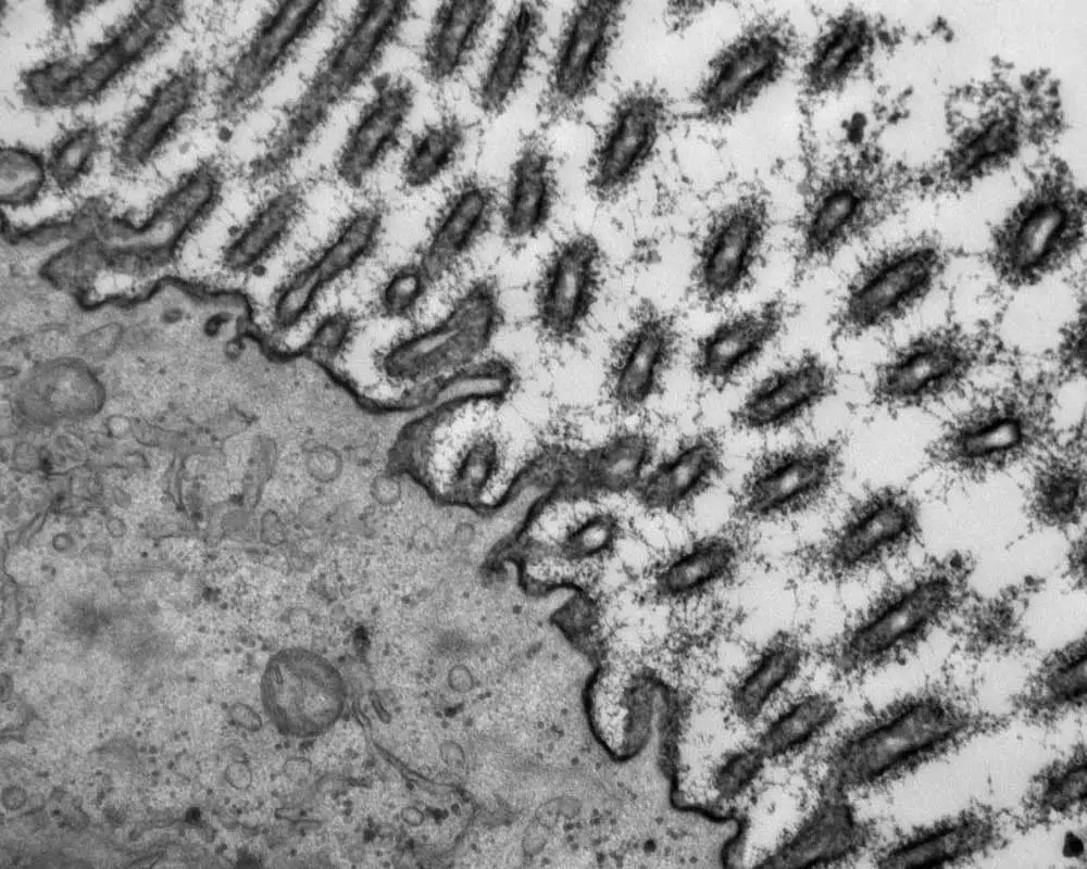
She started off at art school, but that did not quite satisfy her needs. So at 17, her dad took her to visit a laboratory where she saw her first electron microscope. Then in 1960, she saw an advertisement for a technician’s post in a hospital bacteriology lab and took the job, officially starting her science career. To get fully technically qualified, she also needed to take courses in hematology, biochemistry, virology, and histology through employer-sponsored day-release classes.
“I discovered my interest in histology when I saw my first hematoxylin and eosin-stained section of an appendix – that was it. I was completely hooked. I then looked out for jobs and found one in histology that also involved setting up an electron microscopy lab in the Pathology Department of Birmingham University Dental School. That’s where my love of electron microscopy really developed”
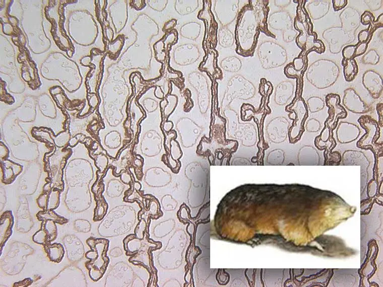
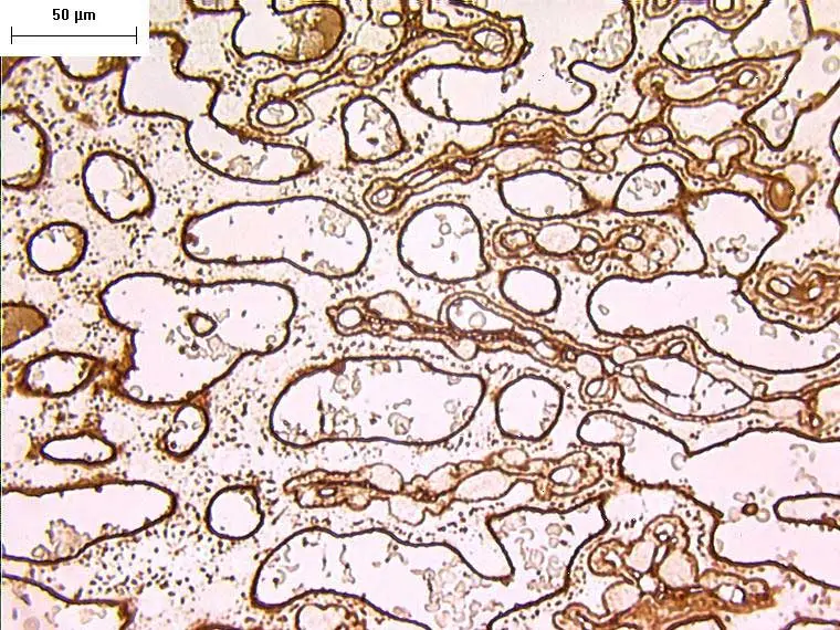
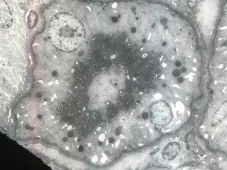
While she thought she was not “clever enough” to go to university, the pathologist she worked with at the Dental School thought differently and encouraged her to apply as a mature student. Having applied to various British Universities, it was Manchester that was particularly interested—if she took just one more advanced class. So, she quit the job, enrolled at a technical college, and completed her secondary level credential. Since then, she has collected a series of degrees, all from the University of Manchester (UK). These range from a BSc (Honors) in Zoology (1970), followed by an MSc on the ultrastructure of rat mammary gland (1971) earned also while working in a pathology lab, and (after an adventure with her husband in Uganda during the Idi Amin years) she was offered a post as research assistant to work on the placenta for her PhD (1976). Most recently, she obtained a Higher Doctorate (DMedSc; 2011), awarded only to candidates who have made original and substantial contributions to their field of study with a body of published work to support their expertise in the field. A workaholic, she continues to write, prolifically, as supported by her lengthy bibliography, which includes 235 full peer-reviewed publications, over 100 abstracts, and 22 book chapters.
“I yearn for those old days of film, but of course, they’ll never come back. You could actually photograph what you saw down through the microscope and you got a really fine, crisp image”
Since her first exposure to electron microscopy in 1964, the instruments have changed significantly, but the biggest change Carolyn notes is the conversion from film to digital imaging. In her days using film, there were 24 pieces of cut film loaded into cassettes in a camera box. A more disciplined (and much slower) approach was needed then, because after exposing one box of film, you would have to stop, empty the cassettes, develop the film, reload new film into cassettes, and wait for the machine to achieve a high vacuum again. While digital technology has the advantage of being able to create hundreds of images non-stop that can all be stored on disk, she sees a distinct disadvantage because of the pixilation artifacts that occur, which were never an issue with film.
“It was just by accident that I got into lectin histochemistry of the animal placenta. I was talking to a Danish veterinary anatomist on a train after a meeting who said that she had some pig placenta. And I said—“Oooh, how interesting—you know I’m working with lectins, and I’d love to do lectin histochemistry on a pig placenta.” She sent me some specimens, and we started collaborating. And then her colleagues offered me cow placenta, sheep placenta, camel and more. This is how my placenta work grew, and I became a one-woman expert on glycosylation of animal placentae.”
When it comes to trying to encapsulate her story-filled science career of over 60 years into a succinct spotlight, one cannot ignore the atypical aspects of a outwardly academic track. She has never had a grant. After a brief post-doctoral position in rheumatology, she was approached by her former Professor in Pathology and offered a permanent position setting up a lectin lab and establishing techniques at the light and electron microscope level. Her post was soon converted to that of Experimental Officer and then to Research Fellow, with no teaching responsibilities and minimal administration, nor did she deal much with students, although now she does help them with their writing. The Pathology Department also ran an electron microscope unit and, following the retirement of its maintenance staff, she unofficially took it over, collaborating with groups both locally and internationally “on the side,” thus funding the running of the machine as well as her research and enabling her to “experiment with lectins and look at whatever tissues I was able to obtain”. Since lectin histochemistry is an inexpensive endeavor where working at microgram concentrations is common, a 5-mg vial can last a long time. That’s important when your love of imaging is visionary.


Stay in the Loop. Join Our Online Community
Together we breakthroughTM
©Vector Laboratories, Inc. 2024 All Rights Reserved.