Click-&-Go® Plus Protein Enrichment Kit
* for capture alkyne-modified proteins *
Product No. 1235
Introduction
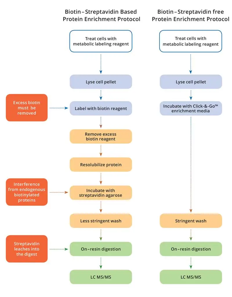
Click-&-Go® Plus Protein Enrichment Kit is an efficient, biotin-streptavidin free tool for capture of alkyne-modified proteins on an picolyl azide-agarose resin. Once covalently attached to the resin via copper catalyzed click chemistry, beads can be washed with highest stringency virtually eliminating any non-specifically bound proteins. Upon protease digestion, this yields a highly pure peptide pool that is ideal for mass spectrometry (e.g., LC MS/MS) based analysis.
Kit Contents
| Component | Amount | Storage |
|---|---|---|
| Picolyl Azide Agarose resin, 50% slurry (Component A) | 2 mL | 2-4°C |
| Lysis buffer (Component B) | 7 mL | 2-4°C |
| Urea (Component C) | 4.8 g | 2-25°C |
| Additive 1 (Component D) | 1.5 mL | 2-4°C |
| Copper (II) Sulfate, 100 mM solution (Component E) | 0.5 mL | 2-25°C |
| Additive 2 (Component F) | 400 mg | 2-25°C |
| Agarose SDS wash buffer (Component G) | 200 mL | 2-4°C |
| Empty spin columns (Component H) | 10 | 2-25°C |
Materials Required but Not Provided for Capturing of Alkyne-tagged Proteins
- 5-20 mg alkyne-tagged cell or tissue extract
- Unlabeled negative control cells or tissue
- Sample rotator
- Table top centrifuge
- Protease Inhibitor
- 2 mL microcentrifuge tubes
- 18 MΩ water
- Probe sonicator
Materials Required but Not Provided for On-resin Digestion with Protease
- DTT
- Iodoacetamide
- 8M Urea/100 mM Tris, pH 8
- Acetonitrile
- Mass-spec grade trypsin
- 0.1% TFA
- C-18 desalting cartridges (e.g. Waters WAT036820)
- Digestion Buffer
- Heat block
- Vacuum Concentrator

Figure 1. Schematic representation of pull-down workflows for biotin-streptavidin and Click-&-Go® enrichment protocols.
Preparation of Stock Solutions
| Lysis Buffer (200 mM Tris pH 8, 4% CHAPS, 1 M NaCl, 8 M Urea) | Add the entire bottle of Solid Urea (Component C) to the bottle containing Lysis Buffer (Component B) provided. Mix the solution on a rotator at room temperature until urea is completely dissolved (1-2 hours). Store refrigerated (for up to 1 week) or at -20°C for 1 year to avoid decomposition of urea. Note: – 30 minutes before starting the enrichment protocol add 20 uL Protease Inhibitor Cocktail (e.g. Sigma 8340) for each 1 mL of Lysis buffer (sufficient for 50-200 million cells or 5-20 mg tissue extract) to be used. |
| Agarose SDS Wash Buffer (100 mM Tris, 1% SDS, 250 mM NaCl, 5 mM EDTA, pH 8.0) | Ready to use. Keep at 2-4°C |
| Additive 2 (Component F) | Add 2 mL of 18 MΩ water to Additive 2 (Component F) and vortex until fully dissolved. After use, store remaining stock solution at -20°C for up to 1 year. |
Protein Enrichment Protocol (per enrichment)
1. Preparation of Picolyl Azide Agarose Resin (Step 1)
- Mix the 50% resin slurry (Component A) until the resin is completely resuspended
- Before the resin settles, transfer 200 µL of well-mixed resin with a 1 mL pipette into a clean 2 mL microfuge tube.
- Add 1.3 mL 18 MΩ water to the resin.
- Pellet the resin by centrifugation for 2 minutes at 1000 x g.
- Carefully aspirate the supernatant leaving approximately 200 µL of settled resin at the bottom of the tube, take care not to aspirate settled resin.
2. Lysate Preparation (Step 2)
- Add 1 mL Lysis Buffer containing Protease Inhibitor (see Preparation of Stock Solutions) to each alkyne-containing cell or tissue extract containing 5-20 mg protein in a 2 mL microfuge tube.
- Incubate the lysis mixture on ice for 5-10 minutes.
- While on ice, sonicate the mixture using a probe sonicator by applying two 3-second pulses. Take care not to overheat the sample.
- Repeat step 2-3 until the lysate is no longer viscous (e.g. viscosity of water).
- Centrifuge the lysate at 10,000 x g for 5 minutes.
- Place lysate back on ice until ready for the click reaction.
3. Preparation of 2X Copper Catalyst Solution (Step 3)
- Prepare 1 mL of 2X Copper Catalyst Solution per enrichment reaction as follows:
860 µL → 18 MΩ water
100 µL → Additive 1 (Component D)
20 µL → Copper (II) Sulfate Solution (Component E)
20 µL → Additive 2 (Component F) - Vortex 2X Copper Catalyst Solution to mix
4. Lysate/Agarose Click Reaction (Step 4)
- Assemble the click reaction in a 2 mL microfuge tube as follows:
200 µL → washed Azide-Agarose resin (Step 1.5)
800 µL → cell or tissue lysate (Step 2.6)
1000 µL → 2X Copper Catalyst Solution (Step 3.2) - Rotate end-over-end on sample rotator for 16-20 hours
5. Reduction & Alkylation of Resin Bound Proteins (Step 5)
- Warm Agarose Wash Buffer w/SDS to room temperature before starting, making sure the solution is homogenous and clear before use.
- Centrifuge agarose resin (Step 4.2) for 1 minute at 1000 x g. Aspirate the supernatant to waste, taking care not to aspirate the resin.
- Add 1.8 mL of 18 MΩ water to the resin, centrifuge at 1000 x g, aspirate the supernatant to waste taking care not to aspirate resin. This water wash step prevents clumping of the resin caused by interaction of residual Lysis Buffer with the SDS in Agarose Wash Buffer.
- Add 1 mL Agarose Wash Buffer w/SDS and 10 μL of 1M DTT to the resin. Vortex briefly to resuspend the resin.
- Heat the resin at 70°C on a heat block for 15 minutes, then cool to room temperature for 15-30 minutes.
- Centrifuge resin for 5 minutes at 1000 x g, aspirate the supernatant to waste taking care not to aspirate the resin
- Prepare 1 mL of a 40 mM iodoacetamide solution per enrichment reaction by dissolving 7.4 mg of iodoacetamide into 1 mL of Agarose Wash Buffer w/SDS.
- Add 1 mL 40 mM iodoacetamide solution to the resin, vortex to resuspend the resin, incubate the reaction in the dark for 30 minutes at room temperature.
6. Resin Wash (Step 6)
Agarose Wash Buffer w/SDS is used for stringent removal of non-specifically bound proteins. After this wash, it is critical to remove residual SDS by washing exhaustively with 8 M urea and 20% acetonitrile prior to mass spectrometry analysis.
- Twist off the spin column’s bottom closure and remove the cap.
- Using a 1 mL pipette resuspend the resin from Step 5.8, then transfer the resin to a spin column.
- Rinse the resin tube with 0.5 mL H2O, and then also transfer this volume to the spin column.
- Add 2 mL of Agarose Wash Buffer W/SDS to the spin column, centrifuge at 1000 x g for 1 minute. Repeat this step 5 times.
- Add 2 mL of 8 M urea/100 mM Tris pH 8 (not provided) to the spin column, centrifuge at 1000 x g for 1 minute. Repeat this step 5-10 times.
- Add 2 mL of 20% acetonitrile (not provided) to the spin column, centrifuge at 1000 x g for 1 minute. Repeat this step 5–10 times.
7. Protease Digestion of Resin-Bound Proteins
- Cap the bottom of the spin column, add 500 μL of digestion buffer (100 mM Tris, 2 mM CaCl2, 10% acetonitrile (not provided) to the resin.
- Using a 1 mL pipette to resuspend the resin in the spin column, then transfer resin to a clean tube.
- Rinse the spin column with 0.5 mL additional digestion buffer, and then add this rinse to the transferred resin.
- Pellet the resin by centrifugation for 5 minutes at 1000 x g. Aspirate the supernatant to waste, leaving approximately 200 μL of digestion buffer in the tube above the resin, taking care not to aspirate the resin.
- Add 10 μL of 0.1 μg/μL trypsin to the resin slurry, gently mix the slurry, then incubate at 37°C for 6 hours to overnight.
8. Preparation of Digest for Mass Spectrometry Analysis
- Pellet the resin from Step 7.5 by centrifugation for 5 minutes at 1000 x g, then carefully transfer the digested supernatant to a clean tube.
- Add 500 μL 18 MΩ water to the resin. Vortex briefly to mix then pellet the resin by centrifugation for 5 minutes at 1000 x g.
- Transfer the rinse supernatant over the resin to the digest supernatant from Step 8.1.
- Add additional 18 MΩ water to the digest to a final volume of 1 mL
Note: this dilutes the acetonitrile concentration to 2%. - Acidify the diluted digest by adding 2 μL of TFA.
- Desalt the digest on a C-18 cartridge using vacuum or gravity flow, allowing each solution to completely flow through the cartridge before adding the next solution
a. Add 1 mL of 50% acetonitrile/0.1% TFA to the cartridge and discard the effluent.
b. Add 1 mL of 0.1% TFA to the cartridge and discard the effluent. Repeat one more time.
c. Add the acidified, diluted digest to the cartridge and discard the effluent.
d. Add 1 mL of 0.1% TFA to the cartridge and discard the effluent. Repeat one more time.
e. Place a clean 1.5 mL tube below the C-18 cartridge outlet.
f. Elute the peptides into a clean 1.5 mL tube by adding 700 μL of 50% acetonitrile/0.1% TFA to the C-18 cartridge. - Dry the eluate containing the desalted peptide digest in a vacuum concentrator. Store at -20°C until ready for MS analysis.
Troubleshooting
| Problem | Possible Cause | Solution |
|---|---|---|
| Low yield of enriched proteins | Inefficient capture or low abundance of azide-tagged proteins | Increase lysate concentration (use more cells) or pre-enrich the proteins (e.g. soluble lysate, membrane lysate, lectin enrichment, etc.). Confirm peptide recovery by measuring A280 after digestion |
| Inefficient digestion of resin-bound proteins | Use high quality trypsin | |
| High background with unlabeled control cells | Insufficient washing of resin | Increase column washes Use only high purity reagents Prepare filtered buffers fresh Ensure proper preparation of copper catalyst solution. |
| Signal suppression during MS analysis | SDS contamination in digest | Wash the resin thoroughly after the Agarose Wash Buffer w/SDS wash with another buffer such as 8M urea and 20% acetonitrile to remove all traces of SDS detergent |

