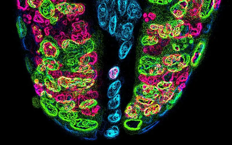
Immunofluorescence (IF) Guide
Immunofluorescence (IF) is a powerful method for visualizing proteins expressed directly within tissues. This method combines immunology and fluorescent molecules to identify localized proteins within defined morphological structures and thus, provide insights into gene expression, protein-protein interactions, and biomarker identification. Whether you’re just starting off doing IF or are a seasoned veteran, this guide will take you through the important steps and considerations needed for all immunofluorescence applications.
This guide offers you,
- An overview of the IF workflow
- Optimization tips for each step
- Selection tips for the most appropriate secondary fluorescent conjugates
- A review of autofluorescence, what it is and how to quench unwanted signal
Please submit the following form to request a copy of this guide. Note that fields marked
with an asterisk * are required.


